|
|
 |
|
Monstrilloida ( Order ) |
|
|
|
Monstrillidae ( Family ) |
|
|
|
Cymbasoma ( Genus ) |
|
|
| |
Cymbasoma bali Desai & Krishnaswamy, 1962 (F,M) | |
| | | | | | | Syn.: | Cymbasoma thompsonii : Dakin & Colefax, 1940. | | | | Ref.: | | | Desai & Krishnaswamy, 1962 (p.163, figs.F,M); Suarez-Morales & Morales-Ramirez, 2003 (p.210: Table 2); Suarez-Morales & McKinnon, 2016 (p.45, figs. F, Rem.) | 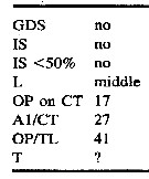 Issued from : E. Suarez-Morales & A. Morales-Ramirez in Proc. Biol. Soc. Washington, 2003, 116 (1). [p.210, Table 2]. Selected taxonomic features in Cymbasoma bali. GDS = anterior half genital double-somite globose; IS = inner seta of P5 smallest; IS <50% = inner seta <50% as long as others; L = longest seta on P5; OP on CT = position of oral papilla on cephalothorax; AI/CT = relative length (as a percent) of A1 to cephalothorax; = relative length (as a percent) of position of oral papilla anteriorly on cephalothorax; T = transverse striations on ''neck'' area. Compare with species of Cymbasoma with uniramous P5 armed with 3 setae: C. concepcionae, C. reticulatum, C. bowmani, C. quintanarooense, C. boxshalli, C. frondipes, C. striatum, C. claparedi, C. tumorifrons.
|
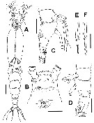
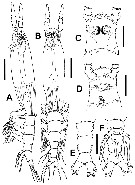 Issued from : E. Suarez-Morales & A.D. McKinnon, in Zootaxa, 2016, 4102 (1). [p.47, Fig.26]. Female (from specimen A): A, habitus (lateral and dorsal, respectively); C, cephalic region (dorsal); D, same (ventral); E, urosome (dorsal); F, same (ventral viewx showing P5). Scale bars: 200µm (A-B); 100 µm (C-F). Nota: Cephalothorax representing 60-61 % of total body length. - Midventral oral papilla noticeably protuberant , located at 19-20 % of cephalothorax length. - Pair of relatively large ocelli present, pigment cups well developed, medially conjointed, separated by less than half an eye diameter, intensely pigmented at inner edges; ventral cup as large as lateral cups. - Frontal area ornamented with pattern of shallow transverse striations and single pair of short frontal sensilla. - Urosome consisting of 5th pedigerous somite, genital double-somite and anal somite, together representing 14-15 % of total body length. - Relative lengths of urosomites 33.8 : 38.4-45.0 : 23-27.8 = 100, respectively. - Genital double-somite longest of urosome. - Anal somite without medial constriction. - Caudal rami weakly divergent, subrectangular, about 1.3 times as long as wide, with 3 caudal setae. - A1 representing about 21.0-21.5 % of total body length and 29-35 % of cephalothorax length, 4-segmented. Relative length of distal antennulary segment 44.8-50 %. - Pedigerous somites 2-4 together accounting for 20 % of total body length. - Ovigerous spines paired,separated at base. Spines slender, about 38-45 % of total body length, straight at their base and along shaft, distally tapering into acute points.
|
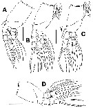
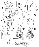 Issued from : E. Suarez-Morales & A.D. McKinnon, in Zootaxa, 2016, 4102 (1). [p.48, Fig.27]. Female (specimen A): A, left A1 (dorsal view); B, urosome (lateral); C, cephalic region (lateral); D, P1; E, P2; F, P3. Scale bars: 50µm (A); 100 µm (B-F). Nota: A1 pattern of setae and aesthetascs see nomemclature from Grygier & Ohtsuka (1995). - P5 medially conjoined at base, bilobate, outer lobe cylindrical, distally truncate and moderately expanded distally. Inner lobe thumb-shaped, noticeably shorter than outer lobe, unarmed, arising proximally from exopodal lobe and reaching about half its length. Exopodal lobe armed with 3 distal setae, innermost seta slightly shorter than the other two.
|
 Issued from : E. Suarez-Morales & A.D. McKinno, in Zootaxa, 2016, 4102 (1). [p.46]. Female: Armature formula of swimming legs P1 to P4. Arabic numerals = setae; Roman numerals = spines. The element (s) on the outer margin of any segment are given first, separted by a hyphen from the inner margin element (s).
|
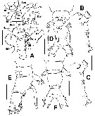
 Issued from : E. Suarez-Morales & A.D. McKinnon, in Zootaxa, 2016, 4102 (1). [p.49, Fig.28]. Female (specimen B): A, habitus (dorsal); B, cephalic tegion (ventral); C, urosome (lateral); D, same (lateral view), specimen C; E, F, distal section of ovigerous spines of two different individuals (B and C). Scale bars: 200 µm (A); 100 µm (B-F)
|
 Issued from : E. Suarez-Morales & A.D. McKinno, in Zootaxa, 2016, 4102 (1). [p.50, Fig.29]. Female (specimen B): A, P1; B, P2; C, P3; D, P4. Scale bars: 100 µm (A-D).
|
 Issued from : E. Suarez-Morales & A.D. McKinno, in Zootaxa, 2016, 4102 (1). [p.51, Fig.30]. Female (specimens B and C): A, left A1 (specimen B); B, cephalic region (lateral); C, same of specimen C; D, urosome and P5 (ventral), specimen C; E, same (lateral); F, same (dorsal). Scale bars: 100 µm (A-F).
|
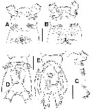 Issued from : E. Suarez-Morales & A.D. McKinno, in Zootaxa, 2016, 4102 (1). [p.52, Fig.31]. Female: A, cephalic region (ventral), specimen D; B, same, specimen C; C, cephalic region (lateral), specimen D; D, urosome and P5 (ventral), specimen A; E, same, specimen C. Scale bars: 100 µm (A-E).
| | | | | Compl. Ref.: | | | Grygier, 1995 a (p.36, 60) | | | | NZ: | 2 | | |
|
Distribution map of Cymbasoma bali by geographical zones
|
| | | | | | | Loc: | | | India (Bombay Harbour), E & SE Australia (Queensland: Davies Reef, Victoria: Port Phillip)
Type locality: Bombay Harbour, India. | | | | N: | 2 | | | | Lg.: | | | (724) F: 1,6; M: 1,5; (1198)* F: 1,3-1,94; {F: 1,60; M: 1,30-1,94}
* Body length measured from the anterior end cephalothorax to the posterior end of the anal somite. | | | | Rem.: | For Suarez-Morales & McKinnon (p.53), the finding of this species in Australian waters represents the first record of this species after its original description more than 50 years ago. based on the resemblance of this species with the illustrated record of females of C. thompsonii from Australia (Dakin & Colefax, 1940), it is suggested that Dakin & Colefax's (1940) record is assignable to C. bali | | | Last update : 19/07/2016 | |
|
|
 Any use of this site for a publication will be mentioned with the following reference : Any use of this site for a publication will be mentioned with the following reference :
Razouls C., Desreumaux N., Kouwenberg J. and de Bovée F., 2005-2026. - Biodiversity of Marine Planktonic Copepods (morphology, geographical distribution and biological data). Sorbonne University, CNRS. Available at http://copepodes.obs-banyuls.fr/en [Accessed March 21, 2026] © copyright 2005-2026 Sorbonne University, CNRS
|
|
|
|


;)
;)
;)
;)
;)
;)
;)

