|
|
 |
|
Monstrilloida ( Order ) |
|
|
|
Monstrillidae ( Family ) |
|
|
|
Caromiobenella ( Genus ) |
|
|
| |
Caromiobenella hamatapex (Grygier & Ohtsuka, 1995) (F) | |
| | | | | | | Syn.: | Monstrilla sp. Sekiguchi, 1982 (p.26, figs.F)
Monstrilla hamatapex Grygier & Ohtsuka, 1995 (p.703, figs.F, N); Chihara & Murano, 1997 (p.1002, Pl.234: F); Ferrari & Dahms, 2007 (p.15, Rem. N); Suarez-Morales, 2011 (p.8, 11); Chang C.Y., 2014 (p.207, Redescr. F, figs F, Rem.; | | | | Ref.: | | | Jeon & al., 2018 (p.47, fig.11, 12, Rem. p.63) | 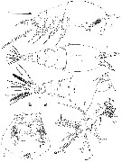 issued from : M.J. Grygier & S. Ohtsuka in J. Crustacean Biol., 1995, 15 (4). [p.710, Fig.5]. As Monstrilla hamatapex. Female (from Tanabe Bay, Japan): A, habitus (lateral; most setae omitted from appendages); B, idem (dorsal; setae omitted from A1); C, anterior end (ventral); D, right A1 (lateral view); E, posterior part of urosome (dorsal); setules omitted from all but 2 of longer caudal setae. Abbreviations (for D, see Fig.6): an = A1 or 1st segment thereof; cg = cerebral ganglion; e, cup of nauplius eye; gs = anterior part of genital double-somite; op = oral papilla; os = ovigerous spines; ps = pit setae; s = simple seta; dc = scars. Scale bars in mm.
|
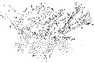 issued from : M.J. Grygier & S. Ohtsuka in J. Crustacean Biol., 1995, 15 (4). [p.712, Fig.6]. As Monstrilla hamatapex. Female: Semischematic diagram of newly devised nomenclature of basic setal armature of A1 in female monstrilloid copepods. it is drawn in dorsal view and a little from the medial side, assuming the antennule is held in its typical orientation, pointing straight forward from the head. There are 4 main kinds of setae: Short, tapered, often spinelike setae (1-6) are arranged more or less medially in clusters of 1-5-1-5-1-2 from proximal to distal; in each group of 5, two are more dorsal (d1-2) and three more ventral (v1-3); Long, setulose, straplike setae (II-V) are associated with the first type in groups of 0-1-2-2-3-0, respectively, and are directed dorsally (d), ventrally (v), or, in one case, medially (m). There are 2 aesthetascs (aes), 1 of them ventral (4aes), the other apical (6aes). There are 6 setae (b) of variable form subapically on the antennule's outer edge; the three more dorsal ones are often highly branched (b1-3); of the others, the middle one may be slightly branched (b5) and two are simple (b4, b6). In some species, like M. hamatapex, none of the b-setae are branched (see Fig.5D
|
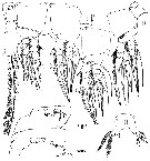 issued from : M.J. Grygier & S. Ohtsuka in J. Crustacean Biol., 1995, 15 (4). [p.713, Fig.7]. As Monstrilla hamatapex. Female: A, right P1 and coupler (posterior view); B, right P2 and coupler (anterior view); ; probable length of broken seta of basis shown as dashed on left leg 2); C, left P3 with short seta on basis (arrow) (posterior view); D, left P4 and coupler (posterior view); E, basis of right P2 (posterior view; enlarged from C, showing sclerite between basis and exopod (arrow)); F, basis of right P2 (anterior view enlarged from B); G, outer apical seta and spinelike seta of exopod of right P4 (posterior view); H, P5 (anterior view, showing aberrant inner seta on right leg. Scale bars in mm.
|
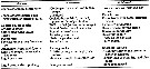 issued from : M.J. Grygier & S. Ohtsuka in J. Crustacean Biol., 1995, 15 (4). [p.718, Table 1]. As Monstrilla hamatapex. Differences between M. hamatapex and European M. helgolandica
|
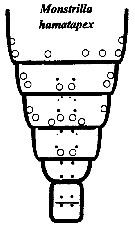 issued from : M.J. Grygier & S. Ohtsuka in Zool. J. Linnean Soc., 2008, 152. [p.499, Fig.29]. As Monstr(illa hamatapex. Female: Dorsal and lateral pore and pit seta patterns, from rear of cephalothorax through genital compound somite. Symbols: dots (three sizes) = pores; larger circles = pits of pit setae. Pattern based on SEM and light microscopical examination.
|
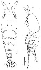 Issued from : C.Y. Chang in Anim. Syst. Evol. Divers., 2014, 30 (3). [p.211, Fig.3]. As Monstrilla hamatapex. Female: A, habitus (dorsal); B, habitus (lateral) . Scale bars: A = 200 µm; B = 300 µm. - Cephalothorax accounting for about 45 % of body length. Dorsal surface ornamented with several sensory pores o, anterior end of cephalothorax, flanking 2 pairs of large pores posteriorly, then with 1 transverse row of 4 small pôres at level of anterior quarter of cephalothorax; 4 pairs of small pores situated middorsally just behind midlength of cephalothorax, these being furnished with subcuticular ducts; 4-5 pairs of sensilla present dorsolaterally near posterior margin of cephalothorax. - Nauplius eye consisting of 1 anteroventral and 2 layrtal small cups, widely separated from each other in dorsal view. - Oral papilla situated slightly anterior to midlength of cephalothorax (42.4 %), slightly protruding ventrally. Weak wrinkles present around oral papilla and nipple-like scars. - Urosome 4-urosomites plus caudal rami, accounting for about 29 % of body length (excluding caudal setae). - Genital double-somite slightly produced outer posteriorly, with transverse wrinkles along posterior margin dorsally. Bearing paired, long ovigerous spines, inserted on middle of ventral surface, basally separated, representing about 43 % of total body length, nearly 1.5 times longer than urosome, with tips pointed, not swollen, extending far beyond tips of caudal setae. - Anal somite trapezoidal; lacking wrinkles or striae both on dorsal and ventral surfaces; lateral margin nearly smooth, without apparent notch. - Caudal rami a little divergent; ramus suboval, with nearly straight inner margin, about 1.3 times than wjde; furnished with 5 long plumose setae, subequal in length and breadth, and 1 slender, short, naked seta dorsal to 2nd medial seta. - A1 short and relatively stubby, about 19 % of total body length, and about 43 % as long as cephalothorax; 2-segmented: 1 short basal segment and 1 long, compound distal segment. Basal segment bearing 1 short naked seta (seta 1) anterodistally. Distal compound segment not clearly defined, with 5 indented parts (possibly representing original segments 2-6, showing standard arrangement of long, plumose setae, 1-2-2-3-0 from proximal to distal). - P5 single-lobed, medial margin with slight swelling representing vestige of endopodal lobe; exopod armed with 2 plumose apical and subapical setae, subequal in length.
|
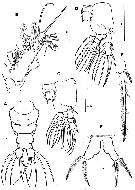 Issued from : C.Y. Chang in Anim. Syst. Evol. Divers., 2014, 30 (3). [p.212, Fig.4]. As Monstrilla hamatapex/.
Female: A, urosome (dorsal); B, A1; C, P1; D, P3; E, outer spine and outer apical seta on 3rd exopodal segment of P3; F, P5.
Scale bars: 100 µm (A-F).
|
 Issued from : C.Y. Chang in Anim. Syst. Evol. Divers., 2014, 30 (3). [p.213]. Female: Armature of swimming legs P1 to P4.
| | | | | NZ: | 1 | | |
|
Distribution map of Caromiobenella hamatapex by geographical zones
|
| | | | Loc: | | | Japan (Tanabe Bay, Ago Bay), Korea (East Sea, South Sea) | | | | N: | 2 | | | | Lg.: | | | (799) F: 2,08; (866) F: 1,5-1,8; (1208) F: 1,321-1,642; {F: 1,32-2,08} | | | | Rem.: | For Jeon, Lee & Soh (2018, p.47, Table 1, 2, fig.11, 12 ) this species belongs to the new genus Caromiobenella, therefore it is a comb. nov., based on the molecular comparison and invertebrate hosts. | | | Last update : 07/01/2025 | |
|
|
 Any use of this site for a publication will be mentioned with the following reference : Any use of this site for a publication will be mentioned with the following reference :
Razouls C., Desreumaux N., Kouwenberg J. and de Bovée F., 2005-2025. - Biodiversity of Marine Planktonic Copepods (morphology, geographical distribution and biological data). Sorbonne University, CNRS. Available at http://copepodes.obs-banyuls.fr/en [Accessed November 16, 2025] © copyright 2005-2025 Sorbonne University, CNRS
|
|
|
|




;)
;)
;)
;)
;)
;)
;)
;)