|
|
 |
|
Platycopioida ( Order ) |
|
|
|
Platycopiidae ( Family ) |
|
|
|
Platycopia ( Genus ) |
|
|
| |
Platycopia orientalis Ohtsuka & Boxshall, 1994 (F,M) | |
| | | | | | | Ref.: | | | Ohtsuka & Boxshall, 1994 (p.152, figs.F,M, juv.); Boxshall & Huys, 1998 (p.768, fig.2); Ferrari & Dahms, 2007 (p.71, Rem.); | 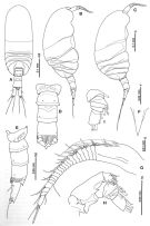 issued from : S. Ohtsuka & G.A. Boxshall in J. Crustacean Biol., 1994, 14 (1). [p.153, Fig.1]. Female (from off Okinawa): A, habitus (dorsal); B, idem lateral right side); C, idem (another specimen); D, urosome (dorsal); E, idem (lateral right side); F, rostrum (frontal view); G, A1; H, A2; I, exopod of A2. Nota : Cephalosome and 1st pedigerous segment incompletely fused, 4th and 5th separated. Urosome 5-segmented. Genital somite small, with minute prominence laterally. 1st to 3rd abdominal somites each fringed with hyaline lamella posteriorly. 3rd abdominal somite longest, with medially incised pseudoperculum largely covering anal somite. Anal somite small, with triangular, ventromedial process on posterior margin. Caudal ramus more than twice as long as wide, having 2 anterolateral setae (setae I and II in Huys & Boxshall, 1991, p.30, 45 : fig.2.1.10C), 1 posterolateral (III), and 3 terminal (IV-VI) setae, and 1 triangular dorsal and 2 ventral terminal processes. Rostrum sharply oiinted, without filaments terminally, with median pore and paired sensilla proximally. A1 22-segmented ; compound 1st segment probably representing fused ancestral segments III-VII, armed with elongate spiniform seta with flagellate tip and dorsal longitudinal ridge. A2: Coxa unarmed ; basis probably incorporating proximal segment of endopod ; free double segment of endopod with 3 medial setae on medial knob representing 2nd rndopodal segment and 7 terminal setae ; exopod indistinctly 7-segmented, setal formula 1, 0, 1, 2, 1, 1, 4 ; 7th segment representing double segment.
|
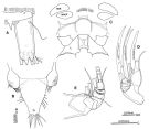 issued from : S. Ohtsuka & G.A. Boxshall in J. Crustacean Biol., 1994, 14 (1). [p.154, Fig.2]. Female: A, caudal ramus (posterior surface); B, labrum; C, position of intermaxillipedal process (Mx = Mx2, L = P1); D, basis and endopod of Mx2; E, endopod of Mxp. Nota : Labrum tripartite ; central lobe strongly produced ventrally, carrying tuft of strong spinules terminally and 2 pores subterminally ; lateral lobes fringed with setules along posterior margin. Intermaxillipedal process not developed, bearingrow of spinules on median sclerite between maxillipeds
|
 issued from : S. Ohtsuka & G.A. Boxshall in J. Crustacean Biol., 1994, 14 (1). [p.155, Fig.3]. Female : A, Md; B, Mx1 (setae on basis and both rami omitted); C, basis and both rami of Mx1; D, Mx2; E, Mxp. Nota : Md : gnathobase with 4 teeth and long setules ; basis elongate, unarmed ; endopod 1-segmented, bearing 6 setae apically ; exopod 4-segmented, setal formula 0, 2, 1, 2. Mx1 : praecoxal arthrite bearing 6 stout spines and 6 setae ; pointed proximal process originating on outer margin of praecoxa ; coxal endite with 3 terminal setae ; coxal epipodite absent ; basal exite small, with 2 small setae terminally ; basal endite elongate with 3 spinulose setae distally ; rami fused with basis distally, with 6 setae along outer margin, representing 2nd basal endite, endopod, and exopod. Mx2 : praecoxal and coxal endites with armature formula 4, 1, 3, 3 ; basal endite with 2 stout and 2 fine setae ; basis and 1st endopodal segment fused to form allobasis, bearing syout endopodal seta distally ; free endopod indistinctly 3-segmented, armature formula 1, 2, 3. Mxp : syncoxa with 2 terminal spinulose setae ; basis bearing 2 medial setae of unequal lengths ; 1st endopodal segment partly incorporatd into basis, with terminal seta directed outward ; 2nd and 3rd segments each having long and short seta ; 4th and 5th each with long seta ; 6th with 4 setae of unequal lengths.
|
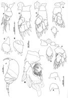 issued from : S. Ohtsuka & G.A. Boxshall in J. Crustacean Biol., 1994, 14 (1). [p.156, Fig.4]. Female: A, P1 (anterior); B, endopod of P1 (anterior); C, exopod of P1 (anterior); D, P2 (anterior); E, P3 (anterior); F, P4 (anterior); ; G, terminal endopod segment of P4 (anterior); H, P5 (posterior); I, endopod of P5 (posterior). Nota: P5 with 2-segmented endopod and 3-segmented exopod; basis lacking inner seta; 2nd segment of endopod and 3rd segment of exopod each with 6 spines. Male: J, habitus (lateral left side); K, 1st endopod segment of P2; L, 1st endopod segment of P4; M, P5 (anterior). Nota: Urosome 5-segmented. A1 15-segmented. P5 symmetrical, biramous; basis with small subterminal outer spine; endopod 3-segmented, 1st segment small, with stout inner spine, 2nd segment with a patch of spinules on distal half surface and inner spine, 3rd segment expanded, covered with numerous spinules distally, and with 4 spines; exopod 3-segmented, 2nd and 3rd segments incompletely fused, inner margin of 3rd segment forming medial hollow, terminal part of segment irregularly expanded, with 4 thick, elongate spines (innermost one of which curved outward approximately at its midlength).
|
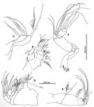
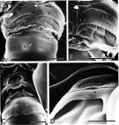 issued from : S. Ohtsuka & G.A. Boxshall in J. Crustacean Biol., 1994, 14 (1). [p.162, Fig.9]. Female (SEM micrographs): A, genital and 1st abdominal somites (ventral view, gonopores indicated by arrows); B, gonopore of genital somite (magnification of A). Male (SEM micrographs): C, genital and 1st abdominal somites (ventral view, gonopores indicated by large arrows, slitlike pore by small arrow); D, slitlike pore of genital somite (magnification of C). Scale = 0.010 mm (A-C, 0.005 mm (D).
|
 issued from : S. Ohtsuka & G.A. Boxshall in J. Crustacean Biol., 1994, 14 (1). [p.158, Fig.6, D]. Male (paratype): D, A1. Nota: A1 15-segmented ; 1st segment compound and bearing large spiniform seta (as in female)
|
 Both A1 male nongeniculate. Sexual dimorphism present in P5. A2 exopod 6-segmented. Exopod of Md indistinctly 3-segmented. Mx2 not extremely modified. Syncoxa of Mxp with 2 setae ...... P1 with 2-segmented exopod and 2-segmented endopod. Exopod of P2 to P5 having inner setae; P2-P5 with outer spines on 1st exopod segment..... Platycopia...... 3 - 1st and 2nd exopod segments of P3 to P5 of female lacking inner setae or spines..... 4 - Endopod of P2 2-segmented...... 5 - Distal 2 endopod segments of female P5 completely fused, bearing 6 spines. Endopod of male P5 3-segmented.
| | | | | Compl. Ref.: | | | Ohtsuka & Nishida, 2017 (p.567) | | | | NZ: | 1 | | |
|
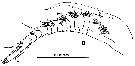
Distribution map of Platycopia orientalis by geographical zones
|
| | | | | |  Chart of 1996 Chart of 1996 | |
| | | | Loc: | | | NW Pacif. ( S Japan: Nansei Islands; off Nagannu Is.: Okinawa) | | | | N: | 1 | | | | Lg.: | | | (602) F: 0,53-0,48; M: 0,44-0,42; {F: 0,48-0,53; M: 0,42-0,44} | | | | Rem.: | epibenthic (depth : 52 m).
For Ohtsuka & Boxshall (1994, p.160) this species is closely related to P. pygmaea Sars in sharing the following apomorphic characters: 1- 1st and 2nd exopod segments of P3-P5 lacking inner setae ofr spines; 2- distal 2nd endopodalal segment of P3 and P4 completely or incompletely fused; 3 - endopod of P2 2-segmented.
The intermaxillipedal process is not developed in P. orientalis, whereas it forms a powerful spinous process in P. inornata and P. perplexa. In the most closely related P. pygmaea, this structure is a slightly raised process bearing a tuft of long, radiating spinules (Huys, pers. comm.). The remarkable development of the intermaxillipedal process appears to be found only in relatively plesiomorphic species of Platycopia. It is likely that the absence of the intermaxillipedal process represents a secondary loss. | | | Last update : 10/05/2019 | |
|
|
 Any use of this site for a publication will be mentioned with the following reference : Any use of this site for a publication will be mentioned with the following reference :
Razouls C., Desreumaux N., Kouwenberg J. and de Bovée F., 2005-2026. - Biodiversity of Marine Planktonic Copepods (morphology, geographical distribution and biological data). Sorbonne University, CNRS. Available at http://copepodes.obs-banyuls.fr/en [Accessed March 26, 2026] © copyright 2005-2026 Sorbonne University, CNRS
|
|
|
|


;)
;)
;)
;)
;)

;)
