|
|
 |
|
Cyclopoida ( Order ) |
|
|
|
Oncaeidae ( Family ) |
|
|
|
Epicalymma ( Genus ) |
|
|
| |
Epicalymma schmitti Heron, 1977 (F,M) | |
| | | | | | | Syn.: | Oncaea schmitti : Malt, 1982 (p.190, figs.F) | | | | Ref.: | | | Heron, 1977 (p.82, figs.F,M); Heron & al., 1984 (p.479, 488, Rem.F); Heron & Frost, 2000 (p.1057, tab.2, 3); Böttger-Schnack & Schnack, 2013 (p.4: Table 1) | 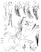 issued from : G.A. Heron in Antarct. Res. Ser. Washington, 1977, 26. [p.85, Fig.28]. Female (SW Pacific-Antarctic area): a, habitus (lateral (scale bar: X); b, posterior of last prosomal segment and urosome (lateral; scale bar: Y); c, urosome (dorsal; scale bar: Y); d, right A1 (scale bar: Y); e, left A2 (scale bar Y); f, labrum (scale bar Z); g, right Md (scale bar: Z); h, right Mx1 (scale bar: Z); i, left Mx2 (scale bar: Z); j, right Mxp (scale bar: Y); k-n, P1 to P4 (scale bar: Y). Nota: Anal segment with operculum margin nearly straight. Urosome (lateral view) with caudal ramus width as wide at midpoint as that of anal segment (posterior to operculum).
|
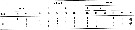 issued from : G.A. Heron in Antarct. Res. Ser. Washington, 1977, 26. [p.84, Table 6]. Female: Armature of swimming legs P1-P4. Si = inner seta; St = terminal seta; Se = outer seta. Arabic numerals: serae; Roman numerals = Spines.
|
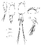 issued from : G.A. Heron in Antarct. Res. Ser. Washington, 1977, 26. [p.87, Fig.29, a-e]. Male: a, habitus (lateral; scald bar: X); b, posterior of last prosomal segment and urosome (lateral; scale bar: Y); c, urosome (dorsal; scale bar: Y); d, left Mxp (scale bar: Y); e, P1 (basis and endopod). Nota: Anal segment with operculum margin straight. Distal corner of last prosomal segment rounded. P5 terminal seta length about same length as that of lateral seta.
|
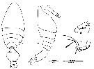 Issued from : S.J. Malt in Bull. Br. Mus. nat. Hist. (Zool), 1982, 42 (3). [p.205, Fig.11, a-d]. As Oncaea schmitti. Female (from 20°N, 21°W); a-b, habitus (dorsal and lateral, respectively); c, A2; d, Mxp. Nota; The specimens are so thinly chitinized that the genital segment had partially collapsed during capture or preservation; therefore, the drawing of the urosome may not represent the morphology of the living animal.
| | | | | Compl. Ref.: | | | Hopkins & Torres, 1988 (tab.1); Razouls & al., 2000 (p.343, tab. 4, 5, Appendix) | | | | NZ: | 5 | | |
|
Distribution map of Epicalymma schmitti by geographical zones
|
| | | | | |  Chart of 1996 Chart of 1996 | |
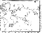 issued from : G.A. Heron & B.W. Frost in Proc. Biol. Soc. Washington, 2000, 113 (4). [p.1061, Fig.25]. issued from : G.A. Heron & B.W. Frost in Proc. Biol. Soc. Washington, 2000, 113 (4). [p.1061, Fig.25].
Locality records for selected species of Epicalymma.
Records include previously published personal observations and data presented in this study (p.1023, Table 3). |
| | | | Loc: | | | Antarct. (Weddell Sea: pack-ice, SW Pacif. SW), off NE Cape Verde Is., Arctic, Norwegian Sea, Station 'P', NE Pacif. | | | | N: | 4 | | | | Lg.: | | | (675) F: 0,72-0,56; M: 0,68-0,52; (679) F: 0,61-0,51; M: 0,51; (680) F: 0,34; 0,33; (848) F: 0,56-0,61; M: 0,52-0,56; {F: 0,33-0,72; M: 0,51-0,68} | | | | Rem.: | bathypelagic, abyssopelagic.
Sampling depth (Antarct.) : 500-1000-2000 m.
For Heron & al. (1984, p.488), the only morphological differences that could be found between specimens from the North and South Polar areas were P2 endopod segments are partially fused on the anterior surface; leg segmentation is often obscure and slightly variable between specimens of a specis. The outer seta on P4 2nd basis is slightly longer on Arctic Ocean females. | | | Last update : 21/12/2016 | |
|
|
 Any use of this site for a publication will be mentioned with the following reference : Any use of this site for a publication will be mentioned with the following reference :
Razouls C., Desreumaux N., Kouwenberg J. and de Bovée F., 2005-2025. - Biodiversity of Marine Planktonic Copepods (morphology, geographical distribution and biological data). Sorbonne University, CNRS. Available at http://copepodes.obs-banyuls.fr/en [Accessed August 17, 2025] © copyright 2005-2025 Sorbonne University, CNRS
|
|
|
|




;)
;)
;)
;)

;)
;)