|
|
 |
|
Calanoida ( Order ) |
|
|
|
Clausocalanoidea ( Superfamily ) |
|
|
|
Aetideidae ( Family ) |
|
|
|
Gaetanus ( Genus ) |
|
|
| |
Gaetanus minutus (Sars, 1907) (F,M) | |
| | | | | | | Syn.: | Chiridius tenuispinus : Campbell, 1929 (p.311);
Gaidius moderatus Tanaka, 1957 a (p.66, figs.F,M);
Gaidius variabilis Brodsky, 1950 (1967) (p.161, figs.F,M); Tanaka & Omori, 1970 (p.127, figs.F); Vaupel-Klein, 1970 (p.12, figs.F,M); Minoda, 1971 (p.24); 1972 (p.326); Deevey & Brooks, 1977 (p.256, tab.2, Station "S"); Gordon & al., 1985 (p.89, Table 2, 3, fish diet); Mackas & Anderson, 1986 (p.115, Table 2); Shih & Marhue, 1991 (tab.2, 3); Hattori, 1991 (tab.1, Appendix); Chihara & Murano, 1997 (p.688, Pl. 45, 46: F,M); Dolganova & al., 1999 (p.13, tab.1); Kosobokova & Hirche, 2000 (p.2029, tab.2); Yamaguchi & Ikeda, 2000 a (p.99); Yamaguchi & al., 2002 (p.1007, tab.1); Yamaguchi & al., 2004 (p.477, tab.2); Ikeda & al., 2006 (p.1791, Table 2); Homma & al., 2011 (p.29, Table 2, 3, abundance, feeding pattern: suspension feeders); Takenaka & al., 2012 (p.1669, fig.2, bioluminescence assay); Abe & al., 2012 (p.100, temporal & vertical changes, gut content)
Gaetanus variabilis : Park, 1975 a (p.13); Yamaguchi & al., 2005 (p.819, Rem. juv, F,M); Yamaguchi & al., 2007 (p.47, vertical distribution, feeding); Takenaka & al., 2012 (p.1669, fig.3, Table 1, bioluminescence assay); Coyle & al., 2014 (p.97, table 3);
Gaidius columbiae Park, 1967 a (p.231, figs.F,M); Harding, 1974 (p.141, tab.2, gut contents); Bradford & Jillett, 1980 (p.58, figs.F,M, Rem., distribution chart); Shih & Marhue, 1991 (tab.3); Takenaka & al., 2012 (p.1669, fig.2, bioluminescence);
Gaidius minutus Sars, 1907 a (p.10, Rem.F); 1925 (p.49, figs.F as G. pusillus ); Farran, 1926 (p.249, Rem.F); Sewell, 1929 (p.100); Rose, 1933 a (p.97, figs.F); Lysholm & al., 1945 (p.13); Sewell, 1948 (p.499, 520, 527); C.B. Wilson, 1950 (p.234); Vervoort, 1952 d (p.3, figs.F); Tanaka, 1957 a (p.64, figs.F); Tanaka, 1965 a (p.18); Furuhashi, 1966 a (p.295, vertical distribution in Oyashio region, Table 6); Park, 1975 a (p.11); Frontier, 1977 a (p.14); Park, 1978 (p.127); Gardner & Szabo, 1982 (p.218, figs.F,M); Wiebe & al., 1988 (tab.7);
Ref. compl.: C.B. Wilson, 1950 (p.234); Grice & Hulsemann, 1967 (p.15); Lapernat, 2000 (comm. pers.); Bradford-Grieve, 2004 (p.284); Ikeda & al., 2006 (p.1791,Table 2); Lewis & al., 2006 (p.501, Table I, fig.F); Homma & Yamaguchi, 2010 (p.965, Table 2); | | | | Ref.: | | | Park, 1975 a (p.13, 30, figs.F,M); Markhaseva & Razzhivin, 1992 (1993) (p.612); Markhaseva, 1996 (p.206, figs.F,M, Notes p.210); Vives & Shmeleva, 2007 (p.582, figs.F,M, Rem.); Lim & al., 2011 (p.42, Redescr.F, figs.F, Rem., molecular sequence) | 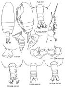 issued from : E.L. Markhaseva in Proc. Zool. Inst. RAN, St. Petersburg, 1996, 268. [p.209, Fig.165]. Female (from different specimens of Kuril Trench): a, b, c ( different localities). Th5 & Gn (lateral view) from K.A. Brodsky after specimens from the NW part of the Pacific Ocean. Nota: Posterior corners with spines of varying size and shape, of the asymmetrical, sometimes covering 1/3 of genital segment length, sometimes reduced to small knobs. A1 longer than cephalothorax reaching the end of genital segment, or the end of caudal rami. Exopodal segments 1 and 2 of P1 incompletely fused (line of fusion visible). P4 coxopodite with 17-20 spines.
|
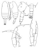 issued from : E.L. Markhaseva in Proc. Zool. Inst. RAN, St. Petersburg, 1996, 268. [p.210, Fig.166]. Male: P5 (a) figure made by K.A. Brodsky after specimens from the north-western part of the Pacific Ocean; other figures from Kuril Trench.
|
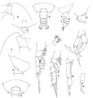 issued from : T. Park in Bull. Mar. Sci., 1975, 25 (1). [p.31, Fig.13]. Female: A, forehead (lateral); B, C, posterior part of metasome and urosome (dorsal and lateral, respectively); D, last metasomal and genital segments (showing a variability of the left metasomal process); E, rostrum (anterior). Male: F, forehead (lateral); G, H, last metasomal and genital segments (lateral and dorsal, respectively); I, distal part of Mx1; J, P1; K, P2; L, P5 (posterior); M, distal part of left P5 (posterior); N, last two exopodal segments of left P5 (posterior). P1-2: legs (anterior), P5: fifth legs. Figures L-N from different specimens. Note: The male is very closely related to that of G. tenuispinus, but can be readily distinguished by the shape of the rostrum and length of the metasomal processes
|
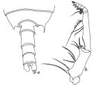 issued from J.C. von Vaupel-Klein in Zool. Verh. Leiden, 1970, 110. [p.13, Fig.2]. As Gaidius variabilis. Female (from NE Pacific: Station \"P\"); b, Mxp. Male: a, dorsal view of the last thoracic somite and urosome.
|
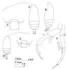 issued from : J.M. Bradford & J.B. Jillett in Mem. N.Z. Oceanogr. Inst., 86, 1980. [p.60, Fig.39]. As Gaidius columbiae. Female: A, habitus (dorsal); B, idem (lateral left side); C, posterior metasome and genital segment (dorsal). Male: D, habitus (dorsal); E, posterior of metasome and genital segment (dorsal); F, P5 (r = right; l = left). Nota: Four of the five male specimens from the soith-west Pacific differ from Park's males in having longer, pointed posterior metasomal extensions, and in the left P5 endopod being shorter.
|
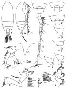 issued from : T. S. Park in J. Fish Bd. Canada, 1967, 24 (2). [p.233, Fig.1]. As Gaidius columbiae. Female: A, habitus (dorsal); B, idem (lateral right side); C-E, posteriorr part of metasome and genital degment (dorsal); F-I, idem (lateral right side); J, rostrum (frontal view); K, A1; L, A2; M, Md; N, Mx1.
|
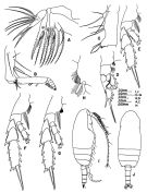 issued from : T. S. Park in J. Fish Bd. Canada, 1967, 24 (2). [p.234, Fig.2]. As Gaidius columbiae. Female: A, Mx2; B, Mxp; C, coxa of Mxp; D, P1; E, P2; F, P3; G, P4; H, inner margin of coxa of P4. Male: I, habitus (lateral right side); J, idem (dorsal). Nota: The two A1 are identical
|
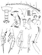 issued from : T. S. Park in J. Fish Bd. Canada, 1967, 24 (2). [p.236, Fig.3]. As Gaidius columbiae. From Strait of Georgia, British Columbia. Male: A, posterior part of metasome and urosome (dorsal); B, rostrum (frontal view); C, A1; D, A2; E, Md (mandibular palp); F, Mx1; G, Mxp; H, P1; I, P4; J, P5 (anterior); K, distal part of left P5.
|
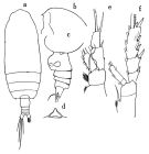 issued from : O. Tanaka in Publ. Seto Mar. Biol. Lab., 1957, VI (1). [p.65, Fig.39]. As Gaidius minutus. Female: a, habitus (dorsal); b, forehead (lateral); c, last thoracic segment and urosome (lateral left side); d, rostrum (anterior aspect); e, P1; f, P2. Nota: Head and 1st thoracic segment, 4th and 5th fused. Last thoracic segment produced into a small nodule on each side. Rostrum short, one-pointed, slightly notched at the apex. Proportional lengths of urosomites and furca 42:13:11:11:23 = 100. First three urosomal segments are fringed with fine teeth on the distal margin. A1 (24-segmented) reaches back to distal end of urosome. Mx1: 9 setae on outer lobe, 11 setae on exopod, 14 setae on endopod, 5 setae on 2nd basal segment, 4 setae on 3rd inner lobe, 14 setae on 1st inner lobe. Mxp: distal inner lobe of 1st basal segment has, beside usual 3 setae, a small lamellous process.
|
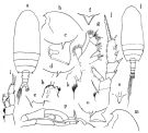 issued from : O. Tanaka in Publ. Seto Mar. Biol. Lab., 1957, VI (1). [p.67, Fig.40]; As Gaidius moderatus. Female: a, habitus (dorsal); b, forehead (lateral); c, last thoracic segment and genital somite (lateral left side); d, idem (other specimen); e, lateral spine of last thoracic segment (dorsal); f, rostrum (anterior aspect); g, Mx1; h, distal margin of 1st basal segment of Mxp; i, P1; j, P2; k, inner margin of 1st basal segment of P4. Nota: Proportional lengths of urosomites and furca 39:17:15:12:17 = 100. A1 (24-segmented) reaches back to distal margin of genital somite. Mx1: outer lobe with 9 setae, exopod with 11 setae, endopod with 14 setae. Male: l, habitus (dorsal); m, forehead (lateral); n, rostrum (anterior aspect); o, last thoracic segment (lateral); p, P5. Nota: Proportional lengths of urosomites and furca 19:29:19:17:2:14 = 100. A1 (20-segmented) extends to distal margin of furca. Mx1: 7 long setae on 1st outer lobe, 11 setae on exopod, 10 setae on endopod.
|
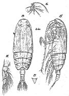 Issued from: G.O. Sars in Résult. Camp. Scient. Prince Albert I, 69, pls.1-127 (1924). [Pl.XIV, figs. 14-18]. Female: 14, habitus (dorsal); 15, idem (lateral left side); 16, forehead (lateral); 17, rostrum (frontal view); 18, P1.
|
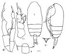 Issued from : K.A. Brodskii in Calanoida of the Far Eastern Seas and Polar Basin of the USSR. Opred. Fauna SSSR, 1950, 35 (Israel Program for Scientific Translations, Jerusalem, 1967) [p.162, Fig.74]. As Gaidius variabilis. Female (from NW Pacif.): habitus (dorsal and lateral left side); R, rostrum; corners of the posterior thoracic segment and genital segment (dorsal); S1, P1. Male: S5, P5.
|
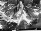 issued from : A.G. Lewis, S. Allen & G. Martens in Crustaceana, 2006, 79 (4). [p.504, Fig.2, A]; As Gaidius minutus. Female (from off British Columbia coast): A, ventral view of the median intersomitic sclerite (MIR) between the maxillipeds base (mxpd-b) and the 1st periopods (P1) rc = ridge crest; TR = transverse sclerite; LIS = lateral intersomitic sclerite
|
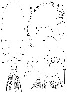 issued from : B.-J. Lim, Song S.J. & Min G.-S. in Korean J. Syst. Zool., 2011, 27 (1). [p.43, Fig.6]. Female (from 36°30'-37°05'N, 131°20'E): A, habitus (dorsal); B, last thoracic segment and urosome (lateral); C-D, rostrum (lateral and ventral, respectively); E, rostrum (enlarged); F, genital segment (ventral); G, anal segment and caudal rami (ventral); H, A1. Scale bars: 1 mm (A-D); 0.25 mm (F, G); 0.5 mm (H). Nota : Prosome about 3.7 times as long as urosome. Cephalosome and 1st pedigerous somite fused. Posterior corners of last pedigerous somite with short spine on each side. Rostrum short and acute, but slightly notched at apex. A1 24-segmented, reaching end of caudal rami.
|
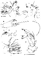 issued from : B.-J. Lim, Song S.J. & Min G.-S. in Korean J. Syst. Zool., 2011, 27 (1). [p.44, Fig.7]. Female: A, A2; B, Md (palp); C, Md (cutting edge of the gnathobase); D, Mx1; E, Mx2; F, Mxp. Scale bars: 0.25 mm (A-C, E, F); 0.01 mm (D). Nota : Endopod of A2 slightly shorter than exopod. Endopod 2-segmented ; exopod 7-segmented. Md : Gnathobase with 6 large and 5 small teeth, 1 long seta and 2 rows of setules. Basis with 2 setae. Endopod 2-segmented, 1st segment with 1 seta, 2nd segment with 9 setae. Exopod 5-segmented, 1st to 4th segments with 1 seta each, distal segment with 2 setae. Mx1 : Arthrite with 4 posterior setae, 9 spines and 1 medial seta. Coxal epipodite with 9 setae. Coxal endite with 4 setae. Basal endtes with 4+5 setae. Endopod with 14 setae. Exopod with 11 setae.
|
 issued from : B.-J. Lim, Song S.J. & Min G.-S. in Korean J. Syst. Zool., 2011, 27 (1). [p.45, Fig.8]. Female: A-D, P1 to P4. Scale bar: 0.025 mm.
|
 issued from : B.-J. Lim, Song S.J. & Min G.-S. in Korean J. Syst. Zool., 2011, 27 (1). [p.45]. Female: Armature formula of swimming legs P1-P4. Roman numerals = spines; Arabic numerals = setae.
|
 Gaetanus minutus female: 1 - Cephalon without frontal spine. 2 - Posterior corners of last thoracic segment with spines of varying size, often asymmetrical. 3 - Exopodal segment 1 of A2 without setae; segment 2 with 2 setae. 2nd internal lobe of Mx1 with 4 setae. Exopod of P1 3-segmented with 2 external spines. 4 - Mxp protopodite without lateral plate. 5 - Md palp base with 2 setae. 6 - Exopod of P1 unclearly 3-segmented.
|
 Gaetanus minutus Gaetanus minutus male: 1 - Cephalon without frontal spine. 2 - Cephalon without crest in anterior part. 3 - Exopodal segment 3 of left P5 stylet-like. Body size (<5 mm). 4 - Posterior corners of last thoracic segment with spines. 5 - Spines significantly shorter than the border of genital segment. 6 - Exopodal segment 3 of P5 with stylet-like narrow. 7 - Body size < 4 mm. 8 - Exopodal segment 2 of left P5 with smooth internal border without excavation near base of exopodal segment 3, segment 2 shorter than segment 3.
|
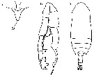 Issued from : M. Chihara & M. Murano in An Illustrated Guide to Marine Plankton in Japan, 1997. [p.714, Pl. 46, fig.47 a-c]. As Gaidius variabilis. Male: a, habitus (dorsal); b, P5; c, rostrum. a, c : after Tanaka, 1957 a; b : after Brodsky, 1950. Numbers show specific characters.
|
 Issued from : M. Chihara & M. Murano in An Illustrated Guide to Marine Plankton in Japan, 1997. [p.713, Pl. 46, fig.45 a-c, d]. As Gaidius variabilis. Female: a, habitus (dorsal); c, rostrum; d, P1. a, d : after Tanaka & Omori, 1970; c : after Tanaka, 1957 a. Numbers show specific characters.
| | | | | Compl. Ref.: | | | Suarez-Morales & Gasca, 1998 a (p108); Sedova & Grigoriev, 2005 (p.112); Kosobokova & al., 2007 (p.929: Tab.7); Galbraith, 2009 (pers. comm.) | | | | NZ: | 10 | | |
|
Distribution map of Gaetanus minutus by geographical zones
|
| | | | | |  Chart of 1996 Chart of 1996 | |
 Issued from : A. Yamaguchi, S. Ohtsuka, K. Hirakawa & T. Ikeda in La mer, 2007, 45. [p.50, Fig.3]. Issued from : A. Yamaguchi, S. Ohtsuka, K. Hirakawa & T. Ikeda in La mer, 2007, 45. [p.50, Fig.3].
Vertical distributions of temperature, salinity, and chlorophyll a (left), total sooplankton biomass and composition of Gaetanus variabilis (middle), and each copepodid stage of G. variabilis (middle to right).
Female/male determination was done for C4 to C6. D: day, N: night.
Nota: Zooplankton samplings were made in the daytime and nighttime with IONESS net (330µm mesh size)through 16 discrete depths between the surface and 1000 m in 26-27 January 1997, at 37°00'N, 131°30'E; 2130 m depth) |
 Issued from : A. Yamaguchi, S. Ohtsuka, K. Hirakawa & T. Ikeda in La mer, 2007, 45. [p.49, Fig.2, a]. Issued from : A. Yamaguchi, S. Ohtsuka, K. Hirakawa & T. Ikeda in La mer, 2007, 45. [p.49, Fig.2, a].
Gaetanus variabilis C6 female. a, Diagram of digestive tract showing three principal zones (1-3). Zones 1-2 were defined as foregut and zone 3 as hindgut in this study. |
 Issued from : A. Yamaguchi, S. Ohtsuka, K. Hirakawa & T. Ikeda in La mer, 2007, 45. [p.49, Fig.2, b-c]. Issued from : A. Yamaguchi, S. Ohtsuka, K. Hirakawa & T. Ikeda in La mer, 2007, 45. [p.49, Fig.2, b-c].
Gaetanus variabilis C6 female. b, Specimenns of which only hindgut filled (gut content score: 3). c, Specimens of which both foregut and hindgut filled (gut content score:4) |
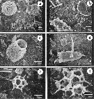 Issued from : A. Yamaguchi, S. Ohtsuka, K. Hirakawa & T. Ikeda in La mer, 2007, 45. [p.53, Fig.5]. Issued from : A. Yamaguchi, S. Ohtsuka, K. Hirakawa & T. Ikeda in La mer, 2007, 45. [p.53, Fig.5].
SEM micrographs of gut of Gaetanus variabilis C6 female.
The diatom Thalassiosira eccebtrica (a, b), tintinnids (Parafavella spp.) (c, d), fragment (hook) of chaetognaths (d), and the silicoflagellate Distephanus speculum (e, f) are seen.
Scale bars indicate 10µm.
Nota: From the morphology of mouthpart appendages, aetid copepods are classified to mixed feeders on both animals and plants. |
 Issued from : J.M. Bradford & J.B. Jillett in New Zealand Ocean. Inst. Memoir, 86, 1980. [p.86, Figs.62]. Issued from : J.M. Bradford & J.B. Jillett in New Zealand Ocean. Inst. Memoir, 86, 1980. [p.86, Figs.62].
Distribution of of Gaidius columbiae (= Gaetanus minutus) in the Tasman Sea and around New Zealand. |
| | | | Loc: | | | off Mauritania, S Azores, Bay of Biscay, Caribbean Sea, G. of Mexico, off Bermuda: Station "S" (32°10'N, 64°30'W), off E Cape Cod, N Atlant., Madagascar (Nosy Bé), W Indian, off S Laccadive Is., E Korea, Japan Sea, Japan, off Sanriku, off E Hokkaido, Station H, Station Knot, Mariana Trench, S Bering Sea, Okhotsk Sea, Kuril-Kamchatka (in Markhaseva & Razzhivin, 1993), off SE Kuril, Bering Sea, S Aleutian Basin, S Aleutian Is., Station "P", British Columbia, Fjord System (Alice Arm & Hastings Arm), Portland Inlet, Guaymas Basin, New Zealand, SW Galapagos (in C. B. Wilson, 1950).
Type locality: NE Atlantic (36°- 46° N, 7°- 27° W) | | | | N: | 43 | | | | Lg.: | | | (1) F: 2,5; (22) F: 3,6-3,2; M: 3,2-3; (37) F: 3,7-2,3; M: 3,4-1,7; (38) F: 2,95; (39) F: 2,62; 3,53; M: 3,19; (113) F: 3,82-3,2; M: 3,65; (201) M: 2,3-1,7; (205) F: 3,3; M, 3,35; (208) F: 3,6-3,25; M: 3,5; (596) F: 3,2-2,97; M: 3,1-2,72; (866) F: 3,2-3,82; M: 3-3,65; (1090) F: 4,46; {F: 2,30-4,46; M: 1,70-3,65} | | | | Rem.: | epi-bathypelagic-? . Sargasso Sea: 1500-2000 m (Deevey & Brooks, 1977, Station "S"); 2000-1000 m (Harding, 1974).
Park (1975 a) and Bradford & al. (1980, p.58) do not agree on the synonymy between this species and G. columbiae.
Markhaseva (1996, p.207) establised the synonymy between this species and G. variabilis Brodsky, 1950.
Yamaguchi & al. (2005, p.819; 2007, p.47) maintain Gaetanus variabilis (Brodsky, 1950), like Abe & al. (2012).
The species is characterized by high variability. | | | Last update : 18/02/2019 | |
|
|
 Any use of this site for a publication will be mentioned with the following reference : Any use of this site for a publication will be mentioned with the following reference :
Razouls C., Desreumaux N., Kouwenberg J. and de Bovée F., 2005-2026. - Biodiversity of Marine Planktonic Copepods (morphology, geographical distribution and biological data). Sorbonne University, CNRS. Available at http://copepodes.obs-banyuls.fr/en [Accessed January 08, 2026] © copyright 2005-2026 Sorbonne University, CNRS
|
|
 |
 |


