|
|
 |
|
Cyclopoida ( Order ) |
|
|
|
Oncaeidae ( Family ) |
|
|
|
Oncaea ( Genus ) |
|
|
| |
Oncaea pumilis Heron, 1977 (F,M) | |
| | | | | | | Ref.: | | | Heron, 1977 (p.75, Descr.F, figs.F); Heron & al.,1984 (p.472, Descr.M, figs.F,M,); Malt & al., 1989 (p.957, Rem.M, figs.M); Böttger-Schnack & Schnack, 2009 (p.131, Table 5: p.140: Rem.); Vives & Shmeleva, 2010 (p.322, figs.F,M, Rem.) | 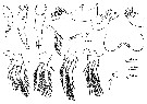 issued from : G.A. Heron in Antarct. Res. Ser. Washington, 1977, 26. [p.76, Fig.23, h-u]. Female (from SW Pacific-Antarctic area): h, habitus (lateral; scale bar: X); i, posterior of last prosomal segment and urosome (lateral; scale bar: Y); j, urosome (dorsal; lacking caudal setae; scale bar: Y); k, left A2 (lacking proximal spine on terminal segment; scale bar: Y); l, labrum (scale bar: Z); m, right Md (scale bar: Z); n, right Mx1 (scale bar: Z); o, right Mx2 (scale bar: Z); p, right Mxp (scale bar: Y); q-t, P1 to P4 (scale bar: Y); u, P5 (scale bar: Z).
|
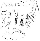 issued from : G.A. Heron, T.S. English & D.M. Damkaer in J. Crustacean Biol., 1984, 4 (3). [p.471, Fig.12, D-J]. Female (from Arctic): D, anal segment and caudal rami (scale bar: w); E, left A2 (scale bar: v). Male: F, habitus (lateral; scale bar: v); G-H, posterior of last prosomal segment and urosome (lateral, dorsal, respectively; scale bar: w); I, left Mxp (scale bar: x); J, P5 (scale bar: x).
| | | | | Compl. Ref.: | | | Razouls & al., 2000 (p.343, tab. 4, Appendix) | | | | NZ: | 3 + 1 doubtful | | |
|
Distribution map of Oncaea pumilis by geographical zones
|
| | | | | | | | | | Loc: | | | Antarctic (SW Pacif.), ? E Medit. (Lebanon), Norwegian Sea, Arctic | | | | N: | 3 | | | | Lg.: | | | (675) F: 0,6-0,5; M: 0,56-0,46; (679) F: 0,52; 0,51; (697) M: 0,48; {F: 0,50-0,60; M: 0,46-0,56} | | | | Rem.: | Sampling depth (Antarct.) : 1000-2000 m.
The geographical distribution of this species is surprising.
The comparison of the male figures by Heron and those by Malt does not appear quite convincing.
For Heron & al. (1984, p.472), the male may be separated from other species by the comparatively slender genital segment (seen in lateral view), and the long anal segment and caudal ramus relative to the preceding orosomal segments. The prosomes of many specimens were abnormally expanded, prsumably deformed by internal cystic growth. | | | Last update : 27/01/2015 | |
|
|
 Any use of this site for a publication will be mentioned with the following reference : Any use of this site for a publication will be mentioned with the following reference :
Razouls C., Desreumaux N., Kouwenberg J. and de Bovée F., 2005-2026. - Biodiversity of Marine Planktonic Copepods (morphology, geographical distribution and biological data). Sorbonne University, CNRS. Available at http://copepodes.obs-banyuls.fr/en [Accessed May 06, 2026] © copyright 2005-2026 Sorbonne University, CNRS
|
|
|
|


;)
;)

