|
|
 |
|
Cyclopoida ( Order ) |
|
|
|
Oncaeidae ( Family ) |
|
|
|
Triconia ( Genus ) |
|
|
| |
Triconia similis (Sars, 1918) (F,M) | |
| | | | | | | Syn.: | Oncaea similis Sars, 1918 (p.193, Descr.F, figs.F); Rose, 1933 a (p.303, figs.F); Jespersen, 1940 (p.93); Sewell, 1948 (p.393, 486); Fagetti, 1962 (p.44); Corral Estrada, 1970 (p. 224, figs.F, Rem.); ? Chen & al.,1974 (p.43, figs.F); ? Ferrari, 1975 (p.224, figs.F); Shuvalov, 1976 (in Kos, 1976, Vol. II, figs.F, Rem.); Heron, 1977 (p.47, figs.F); Dawson & Knatz, 1980 (p.9, 10, figs.F,M); Malt,1983 a (p.6, fig.F, Rem.); ? Malt & al., 1989 (p.957, Descr.M, figs.M); Böttger-Schnack, 1990 (p.869, tab.III); ? Chihara & Murano, 1997 (p.981, Pl.225: F,M); Heron & Frost, 2000 (p.1034, figs.M, tab.2, 3);
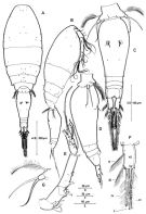
Ref. compl.: Wilson, 1942 a (p.199); 1950 (p.272); De Decker & Mombeck, 1964 (p.13); Shmeleva, 1964 a (p.1068); Mazza, 1966 (p.73); Pavlova, 1966 (p.45); Delalo, 1968 (p.139); Apostolopoulou, 1972 (p.329, 379); Vives, 1982 (p.295); Kovalev & Shmeleva, 1982 (p.86); Sazhina, 1985 a (p.491, tab.3); Greze & al., 1985 (p.8); Lozano Soldevilla & al., 1988 (p.61); Richter, 1994 (tab.4.1a); Böttger-Schnack, 1994 (p.277); Shih & Young, 1995 (p.77); Böttger-Schnack, 1996 (p.1086); 1997 (p.409); Hure & Krsinic, 1998 (p.104); Krsinic, 1998 (p.1051); Hsieh & Chiu, 1998 (tab.2); Razouls & al., 2000 (p.343, Appendix); Oncaea redacta : Heron & Bradford-Grieve, 1995 (p.29, figs.M, no F); Bollens & al., 2011 (p.1358, Table III, fig.6); Selifonova, 2011 a (p.77, Table 1, alien species in Black Sea); Lidvanov & al., 2013 (p.290, Table 2, % composition); Hwang & al., 2014 (p.43, Appendix A: seasonal abundance) | | | | Ref.: | | | Böttger-Schnack, 1999 (p.43, 44, Redescr.F,M, figs.F,M, Rem.: p.52); Krsinic & Grbec, 2002 (p.127, tab.1); Hsieh & al., 2004 (p.398, tab.1, Rem.: p.131); Nishibe & Ikeda, 2004 (p.931, Tab. 2, 4, 5); Böttger-Schnack, 2004 (p.220: tab.2, Rem.); Böttger-Schnack & Schnack, 2009 (p.131, Table 5: Rem.) ; Vives & Shmeleva, 2010 (p.349, figs.F,M, Rem.) | 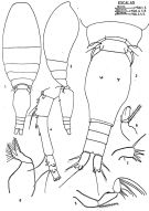 issued from : J. Corral Estrada in Tesis Doct., Univ. Madrid, A-129, Sec. Biologicas, 1970. [Lam.56]. As Oncaea similis. Female (from Canarias Is.): 1, dorsal); 2, idem (lateral right side): 3, urosome (dorsal); 4, A1; 5, A2; 6, Md; 7, Mx2. Nota: The posterior body and furca in the proportional lengths 8:54:9:7:11:11 = 100. A1: the segments in the proportional lengths 9:22:43:12:6:8 = 100
|
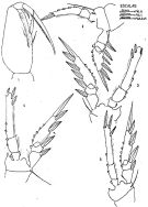 issued from : J. Corral Estrada in Tesis Doct., Univ. Madrid, A-129, Sec. Biologicas, 1970. [Lam.57]. As Oncaea similis. Female: 1, Mxp; 2, P1; 3, P2; 4, P3; 5, P4.
|
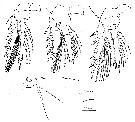
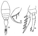 issued from : Q.-c Chen & S.-z. Zhang & C.-s. Zhu in Studia Marina Sinica, 1974, 9. [Pl.7, Figs.14-16]. As Oncaea similis. Doubtful. Female (from China Seas): 14, habitus (dorsal); 15, Mxp; 16, P2.
|
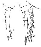 issued from : Q.-c Chen & S.-z. Zhang & C.-s. Zhu in Studia Marina Sinica, 1974, 9. [Pl.8, Figs.1-2]. As Oncaea similis. Doubtful. Female: 1, P3; 2, P4.
|
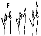
 issued from : R. Böttger-Schnack in Mitt. hamb. zool. Mus. Inst., 1999, 96 [p.46, Fig.2]; Female (from Norwegian fjord): A, habitus (dorsal); B, idem (lateral right side); C, urosome (dorsal); D, idem (lateral left side); E, A1; F, caudal ramus (dorsal, setae are numbered using Roman numerals); G, P5 (dorsal). Nota: Proportional lengths (%) of urosomites and caudal rami 9.7:56.7:5.4:5.4:11.9:10.8. Anal somite 1.4 times wider than long, about same length as caudal rami. Caudal ramus 1.9 times as long as wide. Relative lengths (%) of segments of A1 measured along posterior non-setigerous margin 7.3:23.3:42.2:10.2: 6.3:10.7.
|
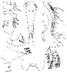
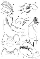 issued from : R. Böttger-Schnack in Mitt. hamb. zool. Mus. Inst., 1999, 96 [p.47, Fig.3]. Female: A, A2 (posterior, lateral elements are numbered using Roman numerals, distal elements numbered using capital letters)); B, labrum (anterior); C, idem (posterior; D, Md (showing individual elements, which are numbered using capital letters); E, Mx1; F, Mx2; G, Mxp (anterior) [g, posteror margin of Mxp basis].
|
 issued from : R. Böttger-Schnack in Mitt. hamb. zool. Mus. Inst., 1999, 96 [p.49, Fig.4]. Female: A, P1 (anterior); B, P2 (anterior); C, P3 (anterior); D, P4 (anterior, intercoxal sclerite omitted).
|
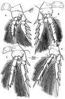
 issued from : R. Böttger-Schnack in Mitt. hamb. zool. Mus. Inst., 1999, 96 [p.51, Fig.5]. Male (from Norwegian fjord): A, habitus (dorsal); B, A1; C, Mxp (posterior) [c, Mxp basis, anterior]; D, urosome (dorsal, spermatophores fully developed); E, idem (ventral); F, idem (lateral left side). Nota: Proportional lengths (%) of urosomites and caudal rami 9.4:58.3:3.8:3.4:3.8:11.6:9.8.
Relative lengths (%) of segments of A1 measured along posterior non-setigerous margin 6.3:25.1:42.4:26.2.
|
 issued from : R. Böttger-Schnack & G.A. Boxshall in J. Plankton Res., 1990, 12 (4) [p.869, Tble III]. Comparison between the females of Oncaea similis (= Triconia similis) species groups. Nota: O; similis; O. minuta; O. dentipes; O. rufa; O. umerus, O. hawii tansfered in the genus Triconia by Böttger-Schnack (1999).
|
 issued from : G.A. Heron & B.W. Frost in Proc. Biol. Soc. Washington, 2000, 113 (4). [p.1033, Fig.9, F-H]. As Oncaea similis. Male (from Sognoforden): F-G, lateral and dorsal, respectively; scale bar: t); H, left Mxp (scale bar: t). Scale bars: 0.1 mm, see figures 3, 4, 5, 10 of Heron and Frost, 2000 for Oncaea canadensis and Oncaea insolita. Nota: Prosome length approximately twice that of urosome. Cephalosome with posterolateral random scatter of minute refractile points associated with pores. Mxp 2nd segment expanded with anterior row of small setules; 2 setae within longitudinal cleft and inner posterior rim with 3 rows of spatulate setules of graduated lengths; terminal claw with narrow process near inner base. Swimming legs as in female. Outer basal seta longer than setae of P5.
|
 issued from : G.A. Heron & B.W. Frost in Proc. Biol. Soc. Washington, 2000, 113 (4). [p.1021, Fig.1, F]. As Oncaea similis. Female: P2-P4 endopod terminal ''spine set'' (combination of the lengths, shapes, and position of the three or two terminal spines on the endopods of swimming legs 2-4 for different Oncaea' species). Nota: P4 with terminal spine less than twice the length of subterminal spine.
|
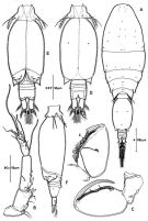
 issued from : G.A. Heron in Antarct. Res. Ser. Washington, 1977, 26. [p.50, Fig.7, d-m]. As Oncaea similis. Female: d, habitus (lateral; scale bar: X); e, urosome (dorsal; lacking caudal setae; scale bar: Y); f, left A1 (scale bar: Y); g, left A2 (scale bar: Y); h, labrum (scale bar: Z); i, right Md (scale bar: Z); j, left Mx1 (scale bar: Z); k, left Mx2 (scale bar: Z); l, right Mxp (scale bar: Y); m, P1 (scale bar: Y). Nota: P5 with free segment small; 2 terminal setae, the longer spiniform and curved posteriorly; seta on body near leg. P6 probably represented by thin spiniform setule near area of external genital apparatus.
|
 issued from : G.A. Heron in Antarct. Res. Ser. Washington, 1977, 26. [p.52, Fig.8, a-d]. As Oncaea similis. Female: a-c, P2 to P4 (scale bar: Y); d, P5 (scale bar: Z).
|
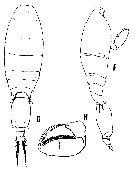
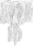 issued from : G.O. Sars in An Account of the Crustacea o Norway, 1918, VI. [Pl. CIX, 1]. as Oncaea similis. Female (from Oster Fjord, near Bergen). ri = endopoditem exp = Mx1.
| | | | | Compl. Ref.: | | | Nishibe & al., 2009 (p.491, Table 1: seasonal abundance); Mazzocchi & Di Capua, 2010 (p.429); Shiganova & al., 2012 (p.61, Table 4); Uysal & Shmeleva, 2012 (p.909, Table I); in CalCOFI regional list (MDO, Nov. 2013; M. Ohman, comm. pers.); Fierro Gonzalvez, 2014 (p.1, Tab. 3, 5, occurrence, abundance); Zakaria & al., 2016 (p.1, Table 1); Benedetti & al., 2016 (p.159, Table I, fig.1, functional characters); | | | | NZ: | 17 | | |
|
Distribution map of Triconia similis by geographical zones
|
| | | | | | | | | | | | | | | | | | | Loc: | | | sub-Antarct. (SW Pacif., Indian), South Africa (E), off S Cape Verde Is., Morocco-Mauritania, Canary Is., G. of Mexico, Hudson Bay, Greenland Sea, Iceland, Norway, North Sea, Medit. (Alboran Sea, W Basin, Ligurian Sea, Tyrrhenian Sea, N & S Adriatic Sea, Aegean Sea, Black Sea, W Egyptian coast, Lebanon Basin), Red Sea, Arabian Sea, Indian (SW, S, subtropical convergence), Philippines, China Seas (East China Sea, South China Sea), off SW Taiwan, Taiwan, Japan (Tosa Bay, Kuroshio & Oyashio regions), Aleutian Is., Station "P", off Washington, Washington inland, off Hawaii, E Pacif., California (San Pedro Bay & Estuary), G. of Panama, off Galapagos, off NW Easter Is., off Chile | | | | N: | 46 | | | | Lg.: | | | (109) F: 0,7-0,67; (133) F: 0,78; (679) F: 0,9-0,84; (681) M: 0,62; 0,63; (687) F: 0,65-0,62; (697) M: 0,54-0,49; (810) F: 0,80-0,72; M: 0,60-0,58; (848) F: 0,74-0,80; M: 0,58-0,68; (864) F: 0,7; (866) F: 0,68-0,59; M: 0,57-0,55; {F: 0,59-0,90; M: 0,49-0,68}
The mean female size is 0.7279 mm (n = 14; SD = 0.0875), and the mean male size is 0.0584 ( n = 10; SD = 0.0527). The size ratio (male : female) is 0.828 (n = 3; SD = 0.0582) or ± 83 %. | | | | Rem.: | Mesopelagic.
Sampling depth (sub-Antarct.) : 0-500-1000 m. According to Hsieh and Chiu (2002) and Hsieh & al. (2004) T. minuta, like T. similis should be indicators of an intrusion of the Kuroshio Current in the East China Sea.
Heron & Frost (2000, p.1017) do not agree that the generic definition of Triconia by Böttger-Schnack (1999) is sufficient to justify inclusion of all of the divergent species she assigned to the new genus.
Could have been confused with other species. Certain locality records are doubtful.
According to Böttger-Schnack (1999, p.53) the species seems have a bipolar distribution rather than being distributed worlwide as shown on the chart. In Chen & al. (1974) the dorsal view of female specimens shows shoulder-like edges between the anterior and posterior part of the genital double-somite, which suggest the specimens belong to T. umerus or T. hawii; the figured distal endopod spine on P2 being as long as the conical process, which is not found in T. similis, but in the closely related T. parasimilis. | | | Last update : 09/12/2020 | |
|
|
 Any use of this site for a publication will be mentioned with the following reference : Any use of this site for a publication will be mentioned with the following reference :
Razouls C., Desreumaux N., Kouwenberg J. and de Bovée F., 2005-2026. - Biodiversity of Marine Planktonic Copepods (morphology, geographical distribution and biological data). Sorbonne University, CNRS. Available at http://copepodes.obs-banyuls.fr/en [Accessed June 16, 2026] © copyright 2005-2026 Sorbonne University, CNRS
|
|
|
|


;)
;)
;)
;)
;)
;)
;)
;)
;)
;)
;)
;)
;)
;)

