|
|
 |
|
Cyclopoida ( Order ) |
|
|
|
Oncaeidae ( Family ) |
|
|
|
Triconia ( Genus ) |
|
|
| |
Triconia umerus (Böttger-Schnack & Boxshall, 1990) (F,M) | |
| | | | | | | Syn.: | Oncaea sp. Böttger-Schnack, 1988, 1990;
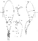
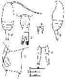
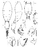
Ocaea umerus Böttger-Schnack & Boxshall, 1990 (p.861, Descr.F, figs.F, tab.III); Böttger-Schnack, 1994 (p.277); 1995 (p.92) 1996 (p.1086); 1997 (p.409); Ohtsuka & al., 1996 a (p.91) | | | | Ref.: | | | Böttger-Schnack, 1999 (p.43, 91, Redescr.F, Descr.M, figs.F,M); Böttger-Schnack & al., 2001 (p.1029, tab.1, 2); Nishibe & Ikeda, 2004 (p.931, Tab. 2, 5); Böttger-Schnack & al., 2004 (p.1130, tab.1, Rem.); Böttger-Schnack, 2004 (p.220: tab.2, Rem.); Di Capua & Boxshall, 2008 (p.1410, figs.F, table 1, 2); Vives & Shmeleva, 2010 (p.351, figs.F,M, Rem.); Böttger-Schnack & Machida, 2011 (p.111, Table 1, 2, fig.2, 3, DNA sequences, phylogeny); Soh & al., 2013 (p.130, figs.F,M) | 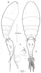 issued from : R. Böttger-Schnack in Mitt. hamb. zool. Mus. Inst., 1999, 96 [p.93, Fig.24]. Female (from Red Sea): A, habitus (dorsal [a, genital aperture and part of genital double-somite, showing surface ornamentation]; B, idem (lateral right side). Nota: Proportional lengths (%) of urosomites and caudal rami 12.8:58.8:7.3:6.4:10.6:11.0. Relative lengths (%) of segments of A1 measured along posterior non-setigerous margin 7:22:45:11:6:9.
|
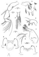 issued from : R. Böttger-Schnack in Mitt. hamb. zool. Mus. Inst., 1999, 96 [p.94, Fig.25). Female: A, A2 (anterior); B, labrum (anterior); C, idem (posterior); D, Md (showing individual elements); E, Mx1; F, Mx2; G, P5 (dorsal); H, caudal ramus (dorsal, terminal setae omitted).
|
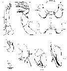
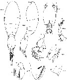
 issued from : R. Böttger-Schnack in Mitt. hamb. zool. Mus. Inst., 1999, 96 [p.96, Fig.26]. Male (from Red Sea): A, habitus (dorsal); B, A1; C, Mxp (anterior); D, urosome (dorsal); E, idem (ventral, spermatophores fully developed); F, idem (lateral right side , spermatophores immature); G, A2 (distal endopod segment). Nota: Proportional lengths (%) of urosomites and caudal rami 11.2:57.4:3.6:3.6:3.6:9.4:11.2. Relative lengths (%) of segments of A1 measured along posterior non-setigerous margin 6.6:24.7:42.3:26.4.
Males were assigned to T. umerus on the length of the terminal accessory seta on caudal ramus, which is longer than the dorsal seta. Males of T. minuta also share this character, but can be distinguished by the length ratio of outer spines on P4 endopd-3, bydifferences in the armature of P5 exopod and by the pore pattern on the dorsal surface of the genital double-somite.
|
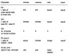 issued from : R. Böttger-Schnack in Mitt. hamb. zool. Mus. Inst., 1999, 96 [p.77, Table 3]. Morphological characters separating males of Triconia minuta, T. umerus, T.rufa and T. hawii. ODS = outer distal spine; OSDS = outer subdistal spine; endp-3 = 3th endopod segment; l = length; sex dimorph. = sexually dimorphic; GS = genital somite; P4, P5 = thoracopods 4, 5; A2 = antenna; CR = caudal rami; lat. arm = lateral armature.
|
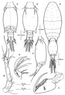
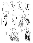
 issued from : J.-H. Wi, K.-S. Shih & H.Y. Soh in Zool. Studies, 2011, 50 (5). [p.596, Fig.5]. Female (from 126°5'E, 32°00N): A-B, habitus (dorsal and lateral, respectively); C, genital double-somite (right side); D, genital aperture.
|
 issued from : J.-H. Wi, K.-S. Shih & H.Y. Soh in Zool. Studies, 2011, 50 (5). [p.597, Fig.6]. Female: A, A1; B, A2; C, labrum (anterior); D, labrum (posterior), arrows indicating round shape of the mid-outer margins of the lobes; E, Md; F, dorsal blade on ghathobase of Md; G, Mx1; H, H, Mxp; I, nasis of Mxp, lacking proximal seta.
|
 issued from : J.-H. Wi, K.-S. Shih & H.Y. Soh in Zool. Studies, 2011, 50 (5). [p.5998, Fig.7]. Female: A-D, P1 to P4, respectively (posterior views).
|
 issued from : J.-H. Wi, K.-S. Shih & H.Y. Soh in Zool. Studies, 2011, 50 (5). [p.602, Fig.9, C]. Female (SEM photograph): C, posterior part of genital double-somite and last part of urosome (dorsolateral view).
|
 issued from : J.-H. Wi, K.-S. Shih & H.Y. Soh in Zool. Studies, 2011, 50 (5). [p.602, Fig.9, D, E]. Female (SEM photographs): D, middle and lateral part of genital double-somite with denticles; E, genital aperture (left side), with a small spinule at the base of the spine
|
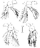
 issued from : J.-H. Wi, K.-S. Shih & H.Y. Soh in Zool. Studies, 2011, 50 (5). [p.600, Fig.8]. Male: A-B, habitus (dorsal and lateral, respectively); C, urosome (ventral); D, P6 and genital flap (right lateral); E, A1; F, A2 (proximal and distal endopodal segments); G, Mxp; H, posterior part of basis of Mxp; I, P2 (distal part of endopodal segment 3).
|
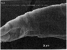
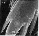 issued from : J.-H. Wi, K.-S. Shih & H.Y. Soh in Zool. Studies, 2011, 50 (5). [p.602, Fig.9, G]. Male (SEM photograph): G, P2 (distal endopodal segment ornamented with denticles).
|
 Issued from : H.Y. Soh, S.Y. Moon & J.H. Wi in Invertebrate Fauna of Korea (eds) Incheon: NIBR, 2013, 21 (28). [p.131, Fig.75]. Female (from Korean waters): A, habitus (dorsal); B, P5 on last thoracic segment and genital segment (lateral);, right side); C, Md; D, Mx1; E, Mxp; F, P2; G, P3; H, P4. Scale bars: A = 200 µm; B, E = 100 µm; C, D = 20Mm; F-H = 20 µm.
|
 Issued from : H.Y. Soh, S.Y. Moon & J.H. Wi in Invertebrate Fauna of Korea (eds) Incheon: NIBR, 2013, 21 (28). [p.132, Fig.76]. Male: A-B, habitus (dorsal and lateral, respectively); C, urosome (dorsal); D, P6 (lateral view); E , A1; F, A2; G, H, Mxp; I, portion of distal endopodal segment of P2. Scale bars: A, B = 100 µm; C, D, F = 50 µm; E, I = 20 µm; G, H = 25 µm.
| | | | | Compl. Ref.: | | | McKinnon & al., 2008 (p.844:Tab.I, p.848: Tab. IV); Nishibe & al., 2009 (p.491, Table 1: seasonal abundance); Böttger-Schnack & Schnack, 2009 (p.131, Table 3, 4); Zenetos & al., 2010 (p.397); Mazzocchi & Di Capua, 2010 (p.429); Tachibana & al., 2013 (p.545, Table 1, seasonal change 2006-2008); Mazzocchi & al., 2014 (p.64, Table 4, 5, abundance); Benedetti & al., 2016 (p.159, Table I, fig.1, functional characters); | | | | NZ: | 6 | | |
|
Distribution map of Triconia umerus by geographical zones
|
| | | | | | | | | | | | | Loc: | | | Medit. (Alboran Sea, Strait of Sicily, Ionian Sea, Lebanon Sea), G. of Aqaba, Red Sea (N-S), G. of Aden, Arabian Sea, Australia (North West Cape), China Seas (East China Sea), Korea, Japan (Tokyo Bay, Tosa Bay, Oyashio region) | | | | N: | 20 | | | | Lg.: | | | (688) F: 0,62-0,57; (810) M: 0,52; (1014) F: 0,580; (1174) F: 0,65-0,71; M: 0,52-0,57; {F: 0,57-0,71; M: 0,52-057} | | | | Rem.: | Mesopelagic. Epipelagic in the Gulf of Naples.
After Wi & al. (2011, p.603) T. umerus and T. denticulata co-occurred south of Cheju Island, where the Kuroshio Current provides the main inflow into the East China Sea and Sea of Japan via the Tsushima Warm Current and Cheju Warm Current.
For Wi & al. (2011, p.599) Triconia umerus from Korean waters agrees with typical morphological characteristics of T. umerus from the Red Sea. However, several form variations exist in the female from Korean waters : 1- the length ratio of the prosome to the urosome (including and exclding caudal rami) is smaller (1.6 :1 and 1.8 :1) compared to Red Sea specimens (2.1 :1 and 2.3 :1) ; 2- the length to the maximum width ratio of the genital double-somite is larger (1.7 :1) than in Red Sea specimens (1.3 :1) ; 3- in the caudal ramus the relative length of seta V to VI is longer (2.6 times), compared to Red Sea specimens (1.8 times) ; 4- Mxp is ornamented with rows of long setules on the anterior surface and rows of setules on the outer margin, not figured in previous descriptions. In the male from Korean waters : 1- the distal endopodal segments of P2-P4 are ornamented with small denticles on the entire surface, but this is not mentioned in the Red Sea specimens ; 2- the length to width ratio of the caudal ramus is larger (1.6 ) than in Red Sea specimens (1.4 times), as in the female ; 3- the proportional length of caudal ramus setae IV and VI is as in the female, but Böttger-Schnack (1999) reported that Rd Sea specimens present a different ratio ; 4- the basis of Mxp is ornamented with several scales distally, and with grooves filled with small spinules on the anterior surface, which were not noted, or erroneously described in previous figures ( the basis lacks scales and grooves, expressed as a patch of spinules) | | | Last update : 21/02/2018 | |
|
|
 Any use of this site for a publication will be mentioned with the following reference : Any use of this site for a publication will be mentioned with the following reference :
Razouls C., Desreumaux N., Kouwenberg J. and de Bovée F., 2005-2026. - Biodiversity of Marine Planktonic Copepods (morphology, geographical distribution and biological data). Sorbonne University, CNRS. Available at http://copepodes.obs-banyuls.fr/en [Accessed June 28, 2026] © copyright 2005-2026 Sorbonne University, CNRS
|
|
|
|


;)
;)
;)
;)
;)
;)
;)
;)
;)
;)
;)
;)
;)

