|
|
 |
|
Cyclopoida ( Order ) |
|
|
|
Paralubbockiidae ( Family ) |
|
|
|
Paralubbockia ( Genus ) |
|
|
| |
Paralubbockia longipedia Boxshall, 1977 (F,M) | |
| | | | | | | Ref.: | | | Boxshall, 1977 a (p.105, figs.F,M); Boxshall & Huys, 1988/89 (p.164, figs.F,M); Huys & Boxshall, 1991 (p.293, figs.F,M); Boxshall & Halsey, 2004 (p.623: figs.F,M) | 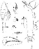 issued from : G.A. Boxshall in Brit. Mus. nat. Hist., Zool., 1977, 31 (3). [p.106, Fig.1]. Female (from 18°N, 25°W): a, habitus (dorsal); b, A2; c, A1 (ventral); d, Md (posterior); e, Mx1 (posterior); f, Mx2 (posterior); g, Mxp (anterior); h, anterior of urosome (ventral). Nota: Prosome longer than urosome. Rostrum small and rounded. Relative lengths orosome somites and caudal rami 15:33:15:12:14:11. Caudal rami about 1.5 times longer than broad. A2 2-segmented. Mxp 4-segmented with a long claw-like distal segment; 2nd segment with a continuous row of spinules along inner margin and 2 naked setae; P5 a free segment with 2 naked apical setae and a lateral seta located on surface of somite near base. P6 probably represented by unarmed cuticularized plate located near genital aperture.
|
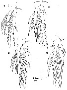 issued from : G.A. Boxshall in Brit. Mus. nat. Hist., Zool., 1977, 31 (3). [p.107, Fig.2]. Female: a-d, P1 to P4 (posterior).
|
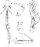 issued from : G.A. Boxshall in Brit. Mus. nat. Hist., Zool., 1977, 31 (3). [p.108, Fig.3]. Male: a, urosome (ventral); b, A1 (ventral); c, A2 (posterior); d, Mx1 (posterior); e, Mx2 (posterior); f, Mxp (anterior). Nota: Urosome 6-segmented; posterior margins of urosome somites 3 to 5 provided with a continuous dentate hyaline frill on ventral surface. Anal somite with a row of fine spinules posteriorly. Caudal rami shorter than anal somite. A1 5-segmented with the distal segment representing the fused 5th to 7th segments of the female.
|
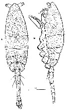 issued from : G.A. Boxshall & R. Huys in Biol. Oceanogr., 1988/1989, 6. [p.165, Fig.1]; Female (from 18°N, 25°W): A-B, habitus (dorsal and lateral, respectively; A1 and swimming legs removed).
|
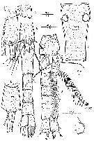 issued from : G.A. Boxshall & R. Huys in Biol. Oceanogr., 1988/1989, 6. [p.166, Fig.2]. Female: A, urosome (ventral); C, anal somite and caudal ramus (dorsal); D, posterior margin of anal somite and caudal ramus (lateral); E, genital double somite (dorsal); F, P6 (lateral). Nota: P5 comprising free distal segment and proximal protopodal segment incorporated into somite; seta on surface of somite representing outer seta of basis; free segment (exopod) about twice as long as wide, armed with a pore, a short inner and long outer seta on distal margin; outer seta sparsely plumose.. Male: B, urosome (ventral). Nota: Urosome 6-segmented, comprising 5th pedigerous somite, genital somite and 4 free abdominal somites.A1 indistinctly 5-segmented (with distal segment representing 3 fused segments of female)
|
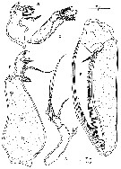 issued from : G.A. Boxshall & R. Huys in Biol. Oceanogr., 1988/1989, 6. [p.167, Fig.3]. Female: A, A2 (posterior); B, Md (anterior); C, Mx1 (ventral; wiyth scar left by missing element indicated by arrow); D, Mx2 (posterior); E, Mxp (lateral).
|
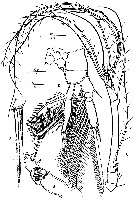 issued from : G.A. Boxshall & R. Huys in Biol. Oceanogr., 1988/1989, 6. [p.169, Fig.5]. Female: A, A1 (dorsal); B, Mx1 (ventral); C, P3 (anterior; outer basal seta missing); D, P3 (detail of tip of endopod).
|
 issued from : G.A. Boxshall & R. Huys in Biol. Oceanogr., 1988/1989, 6. [p.167]. Female & Male: Armature of swimming legs P1 to P4. Roman numeral = spine; arabic numeral = seta.
|
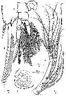 issued from : G.A. Boxshall & R. Huys in Biol. Oceanogr., 1988/1989, 6. [p.168, Fig.4]. Male: A, A1 (ventral); B, labrum (ventral view); C, P1 (anterior); D, P1 (detail of distal part of exopod segment 3). Nota: Labrum: a small flattened plate, unarmed but with a large apical pore.
|
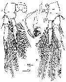 issued from : G.A. Boxshall & R. Huys in Biol. Oceanogr., 1988/1989, 6. [p.170, Fig.6]. Female & Male: A, P2 (anterior); B, P4 (anterior); C, Md (of second female paratype showing variation in spinulation pattern.
|
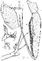 issued from : G.A. Boxshall & R. Huys in Biol. Oceanogr., 1988/1989, 6. [p.171, Fig.7]. Male: A, A2 (lateral); B, Md (anterior); C, Mx1 (ventral); D, Mx2 (possibly incomplete); E, Mxp (lateral). Nota: Claw of Mxp apparently representing an endopodal segment plus terminal claw, with trace of suture between them; inner margin of incorporated endopodal segment bearing a row of spinules and an apical seta. The reduction in the Md and in the praecoxal endite of Mx1 suggests that the adult male is nonfeeding. This is quite a common strategy among planktonic \"poecilostomatoids\", also found in males of the sapphirinid genus Copilia for example. In both sexes the palp ot Mx1 is separated from the proximal segment by a complete structure. This is another unique plesiomorphy within the \"Poecilostomatoid\", since Mx1 is typically reduced to a bilobed orocess in \"Poecilostomatoids\".
| | | | | NZ: | 1 | | |
|
Distribution map of Paralubbockia longipedia by geographical zones
|
| | | | Loc: | | | NE Atlant. (off N Cape Verde Is.) | | | | N: | 2 | | | | Lg.: | | | (139) F: 1,43-1,19; M: 1,19; {F: 1,19-1,43; M: 1,19} | | | | Rem.: | meso-bathypelagic. | | | Last update : 27/01/2015 | |
|
|
 Any use of this site for a publication will be mentioned with the following reference : Any use of this site for a publication will be mentioned with the following reference :
Razouls C., Desreumaux N., Kouwenberg J. and de Bovée F., 2005-2026. - Biodiversity of Marine Planktonic Copepods (morphology, geographical distribution and biological data). Sorbonne University, CNRS. Available at http://copepodes.obs-banyuls.fr/en [Accessed February 20, 2026] © copyright 2005-2026 Sorbonne University, CNRS
|
|
 |
 |


