|
|
 |
|
Calanoida ( Order ) |
|
|
|
Diaptomoidea ( Superfamily ) |
|
|
|
Pseudodiaptomidae ( Family ) |
|
|
|
Pseudodiaptomus ( Genus ) |
|
|
| |
Pseudodiaptomus trihamatus Wright, 1937 (F,M) | |
| | | | | | | Syn.: | Mazellina galleti (F) Rose, 1957 (p.235, figs.F, no M);
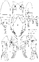
Pseudodiaptomus penicillatus Li & Huang, 1984 (p.386, figs.), after Walter & al., 2006 (p.203, 211, Rem.) | | | | Ref.: | | | Wright, 1937 a (p.155, Descr.M, fig.M); Sewell, 1948 (p.421); Grindley, 1984 (p.221, Rem.); Walter, 1984 (p.380, Descr.F,M, figs.F,M; Rem.); 1986 (p.133); 1986 a (p.503); 1987 (p.367); Chihara & Murano, 1997 (p.894, Pl.164: F,M); Walter & al., 2002 (p.650, fig.F, Table 3); Medeiros & al., 2006 (p.241, figs.M, Table 1, Rem.); Walter & al., 2006 (p.203, 211, Rem.) | 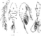 issued from : M. Rose in Bull. Mus. Hist. Nat., 1957, 2ème ser., XXIX (2). [p.236, Fig.1]. As Mazellina Galleti. Female (from Nha-trang, Viet Nam): A1, antennule; D, habitus (dorsal); L, idem (lateral right side); Abd, urosome (lett side view).
|
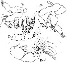 issued from : M. Rose in Bull. Mus. Hist. Nat., 1957, 2ème ser., XXIX (2). [p.237, Fig.2]. As Mazellina Galleti. Female: Md, mandible; A2 antenna; Mxp1, maxilla; Mxp2, maxilliped.
|
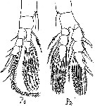 issued from : M. Rose in Bull. Mus. Hist. Nat., 1957, 2ème ser., XXIX (2). [p.239, Fig.3]. As Mazellina Galleti. Female:3, swimming legs P1 and P2.
|
 issued from : M. Rose in Bull. Mus. Hist. Nat., 1957, 2ème ser., XXIX (2). [p.240, Fig.3 bis]. As Mazellina Galleti. Female: 3 bis, swimming legs P3 and P5.
|
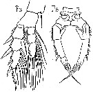
 issued from : T.C. Walter in Proc. Biol . Soc. Wash., 1984, 97 (2). [p.382, Fig.5]. Female (from Philippines): A, habitus (dorsal); B-C, last thoracic segment and urosome (lateral right and left sides, respectively); D, P5 (posterior view). Male: E, habitus (dorsal); F, urosomal segments 1-3 showing spinule rows (ventral); G, last thoracic segment and urosome (lateral left side); H, P5 (posterior view); I, idem (anterior view).
|
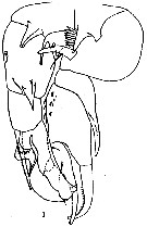 issued from : S. Wright in Ann. Acad. Brasileira Sci., 1937, 9 (2). [Plate 1, Fig.1]. Male (from Luzon, Philippines): 1, P5. Nota: Rostrum bifid, long, slender. Head and 1st thoracic segment fused, line of separation between thoracic segments 4 and 5 present but not conspicuous; segment 5 with single spine, other thoracic segments without spines. urosomite segments 2, 3, 4 with spinules on posterior borders. P5 massive, legs close together; right slightly longer than left; right basipod 1 small, with a large triangular process on inner border; basipod 2 very large, greatest length and breadth almost equal, with 3 notable structures, all rising near the inner border and directed toward the median line: (1) a curved digitiform process with a short and long spine; (2) a cluster of knife-blade spinesq, and (3) a process with 3 salient points which are directed ventrally. Exopodite displaced into medial line; right exopodal segment 1 articulated at inner angle of basipod 2, small, with lateral process which extends distally several times the length of the segment proper; right exopodal segment 2 large, outer distal angle produced distally, lateral spine located at outer distal angle, long, curved inward, reaching to end of terminal hook; on inner border and anterior face of the right exopodal segment 2, several small spines; on posterior face near inner border, 3 small semi-lunar lamellae; right exopodal segment 3 (terminal hook) articulated at inner distal angle of right exopodal segment 2, somewhat shorter than right exopodal segment 1, gently curved, base slightly bulbous bearing slender spine and short digitiform process with delicate spine at the apex. Endopd lacking. Left basipodal segment 1 larger than right basipodal segment 1; posterior face with large process in form of bird's bill; anterior face with a row of large knife-blade spines; left basipodal segment 2 large, length greater than breadth, with a large acuminate spine and 3 hairs on posterior face; left exopodal segment 2 not large, nearly square; length of spine at outer distal angle twice greatest breadth, outer half hyaline, outer border sinuate, near border a semi-lunar serrate structure, segmet bears 3 delicate spines and a sigmoid ridge. Endopodte lacking. Left foot somewhat shorter than the right.
|
 issued from : T.C. Walter, S. Ohtsuka, S. Putchakarn, K. Pinkaew & S. Chullasorn in Proc. Biol. Soc. Washington, 2002, 115 (3). [p.665, Fig.10, A]. Female: A, genital double somite and 2nd urosomal segment (ventral view) Scale bar: 0.01 mm.
| | | | | Compl. Ref.: | | | Lo & al., 2004 (p.468, tab.2): Ohtsuka & al., 2008 (p.115, Table 5); Almeida LR. & al., 2012 (p.13, Table 1, abundance) | | | | NZ: | 4 | | |
|
Distribution map of Pseudodiaptomus trihamatus by geographical zones
|
| | | | | | | Loc: | | | Viet Nam, Celebes, Philippines (Padre Burgos), China Seas (East China Sea), Taiwan (Tapong Bay), Japan, SW Atlant. (N Brazil, Guarairas Lagoon) | | | | N: | 7 | | | | Lg.: | | | (866) F: 1,18-1,28; M: 0,94-1; (998) F: 1,2; {F: 1,18-1,28; M: 0,94-1,0} | | | | Rem.: | freshwater-brackish.
This species was first encountered in shrimp ponds along the margins of the Potengi River estuary (Natal). Its presence was attribuated to an accidental introduction from a breeder stock of the shrimp Penaeus monodon Fabricius, 1798, imported from Philippines.
In Hyalinus species group (trihamatus subgroup) after Walter & al. 2006, p.203. | | | Last update : 04/07/2015 | |
|
|
 Any use of this site for a publication will be mentioned with the following reference : Any use of this site for a publication will be mentioned with the following reference :
Razouls C., Desreumaux N., Kouwenberg J. and de Bovée F., 2005-2026. - Biodiversity of Marine Planktonic Copepods (morphology, geographical distribution and biological data). Sorbonne University, CNRS. Available at http://copepodes.obs-banyuls.fr/en [Accessed June 21, 2026] © copyright 2005-2026 Sorbonne University, CNRS
|
|
|
|


;)
;)
;)
;)
;)
;)
;)

