|
|
 |
|
Calanoida ( Order ) |
|
|
|
Clausocalanoidea ( Superfamily ) |
|
|
|
Scolecitrichidae ( Family ) |
|
|
|
Macandrewella ( Genus ) |
|
|
| |
Macandrewella stygiana Ohtsuka, Nishida & Nakaguchi, 2002 (F,M) | |
| | | | | | | Ref.: | | | Ohtsuka & al., 2002 (p.534, figs.F,M) | 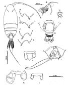 issued from : S. Ohtsuka, S. Nishida & K. Nakaguchi inJ. Nat. Hist., 2002, 36 [p.535, Fig.2]. Female (from off Tokashiki Shima Is.): A, habitus (dorsal); B, rostrum (lateral); C, idem (ventral); D-E, prosomal end (lateral right side, from different specimens); F-G, idem (lateral left side, from different specimens); H, urosome (dorsal); I, P5 and genital double-somite with spermatophore (ventral); J-L, P5 (anterior, from different specimens). Holotype: A-D, F, H-I; paratypes: E, G, K, L.
|
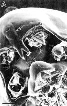 issued from : S. Ohtsuka, S. Nishida & K. Nakaguchi inJ. Nat. Hist., 2002, 36 [p.536, Fig.3]. Female (SEM micrographs): A, rostrum and labrum (ventral, cuticular lens indicated by arrow).
|
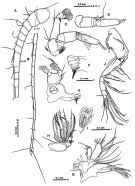 issued from : S. Ohtsuka, S. Nishida & K. Nakaguchi inJ. Nat. Hist., 2002, 36 [p.538, Fig.5]. Female: A, A1 (segments I to XV); B, A1 (segments XVI to XXVII-XXVIII); C, A2; D, exopod of A2; E, Md; F, Md (mandibular cutting edge); G, Mx1; H, Mx2; I, endopod of Mx2; J, Mxp.
|
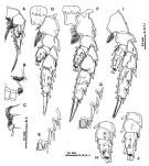 issued from : S. Ohtsuka, S. Nishida & K. Nakaguchi inJ. Nat. Hist., 2002, 36 [p.539, Fig.6]. Female: A, P1 (anterior); B, outer distal margin of endopod of P1 (anterior); C, inner margin of 1st and 2nd exopodal segments of P1; D, P2 (anterior); E, outer margin of 3rd exopodal segments of P2 (anterior); F, P3 (anterior); G, outer margin of 3rd exopodal segment of P3 (anterior); H, endopod of P3 (anterior); I, P4 (anterior); J, endopod of P4 (anterior).
|
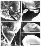 issued from : S. Ohtsuka, S. Nishida & K. Nakaguchi inJ. Nat. Hist., 2002, 36 [p.537, Fig.4]. Female: A, prosomal ends and P5 (indicated by arrow), ventral; B, dorsolateral process of prosomal end (lateral); C, P5; D, terminal portion of right P5; E, terminal portion of left P5. Scales: 0.1 mm (A); 0.05 mm (B-C); 0.01 mm (D-E).
|
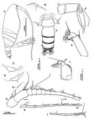 issued from : S. Ohtsuka, S. Nishida & K. Nakaguchi inJ. Nat. Hist., 2002, 36 [p.540, Fig.7]. Male: A, habitus (lateral right side); B, rostrum (lateral); C, prosomal end (lateral right side); D-E, prosomal end (lateral left side); F, urosome (dorsal); G, A1 (segments I to X-XV); H, A1 (segments XVI-XVII to XXII); I, A1 (segments XXIII to XXVII-XXVIII); J, exopod of A2 (seta indicated by arrow more developed in male than in female); K, endopod of A2 (seta indicated by arrow shorter in male than in female); L, Md (mandibular basis and endopod, setae arrowed shorter in male than in female).
|
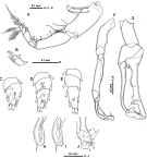 issued from : S. Ohtsuka, S. Nishida & K. Nakaguchi inJ. Nat. Hist., 2002, 36 [p.541, Fig.8]. Male: A, Mxp (with abnormal endopod); B, normal terminal endopodal segments of Mxp; C,
|
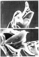 issued from : S. Ohtsuka, S. Nishida & K. Nakaguchi inJ. Nat. Hist., 2002, 36 [p.542, Fig.9]. Male (SEM micrographs): A-B, terminal portion of exopod of left P5 (elements indicated \"a - d\"). Compare with M. omorii). Scales: 0.05 mm.
|
 Issued from : S. Ohtsuka, S. Nishida & K. Nakaguchi in J. Nat. Hist., 2002, 36. [p.543, Table 2]. Seta and spine formula of swimming legs P1 to P4 in female or male..
| | | | | NZ: | 1 | | |
|
Distribution map of Macandrewella stygiana by geographical zones
|
| | | | | | | Loc: | | | NW Pacif. (SW Okinawa, Tokashiki Shima) | | | | N: | 1 | | | | Lg.: | | | (903) F: 3,23-3,84; M: 3,25-3,81; {F: 3,23-3,84; M: 3,25-3,81} | | | | Rem.: | hyperbenthic (Depths: 95-167 m).
For Ohtsuka & al. (2002, p.544) this species is closely related to M. cochinensis from off Cochin (Indian Ocean), on the basis of the structure of the male P5. It is also similar to that of M. joanae. | | | Last update : 06/04/2016 | |
|
|
 Any use of this site for a publication will be mentioned with the following reference : Any use of this site for a publication will be mentioned with the following reference :
Razouls C., Desreumaux N., Kouwenberg J. and de Bovée F., 2005-2026. - Biodiversity of Marine Planktonic Copepods (morphology, geographical distribution and biological data). Sorbonne University, CNRS. Available at http://copepodes.obs-banyuls.fr/en [Accessed February 11, 2026] © copyright 2005-2026 Sorbonne University, CNRS
|
|
 |
 |


