|
|
 |
|
Harpacticoida ( Order ) |
|
|
|
Cervinioidea ( Superfamily ) |
|
|
|
Aegisthidae ( Family ) |
|
|
|
Nudivorax ( Genus ) |
|
|
| |
Nudivorax todai Lee & Huys, 2000 (F,M) | |
| | | | | | | Ref.: | | | Lee & Huys, 2000 (p.5, figs.F,M) | 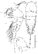 issued from : W. Lee & R. Huys in Zool. J. Linnean Soc., 2000, 129. [p.7, Fig.2]. Female (from Sagami Bay, SE Hatsushima Island): A, habitus (dorsal); B, idem (lateral; arrow indicating folded membranous integument); C, cephalosome and 1st pediger somite (lateral; in telescoped condition, arrowed). Scale in microns. Nota: Prosome 5-segmented, comprising cephalosome and 4 free pediger somites; without surface reticulation; ornamentation consisting of pores and few sensillae, pediger somite 1 without sensillae. Urosome 5-segmented, comprising pediger somite 5, genital double-somite and 3 free abdominal somites; all urosomites with pattern of surface ornamentation consisting of small spinules or denticles dorsally and ventrally. Anal somite with large anal opening; anal operculum vestigial, bordered by tiny spinules anteriorly.
|
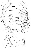 issued from : W. Lee & R. Huys in Zool. J. Linnean Soc., 2000, 129. [p.9, Fig.3]. Female: A, A1 (dorsal); B, Md; C, Mxp; D, anterior margin ao segment 3 of A1 (ventral).. Scale in microns. Nota: A1 7-segmented, without spinous processes on anterior margin of segment 2
|
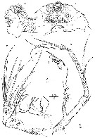 issued from : W. Lee & R. Huys in Zool. J. Linnean Soc., 2000, 129. [p.10, Fig.4]. Female. A, A2; B, detail of distal armature of A2 endopod; C, labrum (posterior); D, left paragnath (anterior); E, Mx2. Scale in microns. Nota: A2 with indistinctly 3-segmented exopod (armature formula 1-1-1); endopod with 3 lateral and 6 apical elements.
|
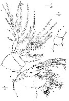 issued from : W. Lee & R. Huys in Zool. J. Linnean Soc., 2000, 129. [p.11, Fig.5]. Female. A, P1; B, Mx1; C, P1 endopod (posterior). Scale in microns.
|
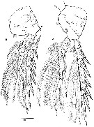 issued from : W. Lee & R. Huys in Zool. J. Linnean Soc., 2000, 129. [p.13, Fig.6]. Female. A, P3 (anterior); B, P2 (anterior; outer basal seta arrowed). Scale in microns.
|
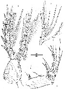 issued from : W. Lee & R. Huys in Zool. J. Linnean Soc., 2000, 129. [p.14, Fig.7]. Female. A, P4 (anterior) Male: B, P4 endopod; C, Mx2 (arrow indicating rudimentary seta on posterior surface). Scale in microns.
|
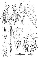 issued from : W. Lee & R. Huys in Zool. J. Linnean Soc., 2000, 129. [p.15, Fig.8]. Female. A, urosome (ventral); B, idem (lateral); idem (dorsal). E, refion around seta I; F, seta II; G, distal margin of caudal ramus (dorsal); H, distal part of P5 (posterior). Male: D, P5 (anterior). Scale in microns. Nota: Each caudal ramus with 7 setae.
|
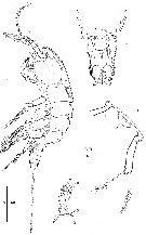 issued from : W. Lee & R. Huys in Zool. J. Linnean Soc., 2000, 129. [p.16, Fig.9]. Female: D, A2 exopod. Male. A, habitus (lateral); B, penultimate and anal somites (dorsal); C, genital field (ventral; minute copulary pore arrowed); D, A2 exopod Scale in microns.
|
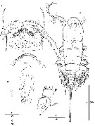 issued from : W. Lee & R. Huys in Zool. J. Linnean Soc., 2000, 129. [p.17, Fig.10]. Male: A, habitus (dorsal); B, anterior area of cephalosome (dorsal); C, genital and 1st abdominal somites (vental; arrw indicating inner vestigial seta); D, Md. Scale in microns. Nota: Cephalosome with linear array of pores along anterior margin. P5 indistinctly 2-segmented, basis and exopod 1 partly fused; exopod2 with 2 outer spines, 2 apical spines and 2 inner setae. P6 with 1 vestigial and 2 well developed setae, medial margin with 2 spinular tufts.
|
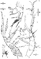 issued from : W. Lee & R. Huys in Zool. J. Linnean Soc., 2000, 129. [p.19, Fig.11]. Male: A, A1 (dorsal); B, Mx1 (posterior); C, Mxp (membranous inserts marking original segmentation arrowed); D, segments 3-4 of A1 (anterior); E, distal half of segment 5 of A1 (anterior). Scale in microns. Nota: A1 9-segmented, geniculation between segments 7 and 8.
|
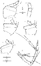 issued from : W. Lee & R. Huys in Zool. J. Linnean Soc., 2000, 129. [p.20, Fig.12]. Male: A, A2; B, protopod and proximal exopod segment of P2; C, idem of P3; D, idem of P4; E, protopod and endopod of P1 (posterior); F, P1 endopod (posterior).. Scale in microns.
|
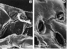 issued from : W. Lee & R. Huys in Zool. J. Linnean Soc., 2000, 129. [p.21, Fig.13, A-B]. Male: A, lateral view of cephalosome showing pores; B, detail of cephalosome pores. Scale bar in microns: A = 20; B = 5.
|
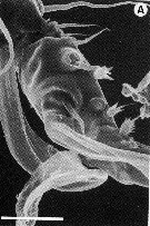 issued from : W. Lee & R. Huys in Zool. J. Linnean Soc., 2000, 129. [p.24, Fig.15, A]. Male: A, A1 (anterior view of segments 4-6). Scale bar in microns = 20.
| | | | | NZ: | 1 | | |
|
Distribution map of Nudivorax todai by geographical zones
|
| | | | Loc: | | | Japan (SE Hatsushima Is.) | | | | N: | 1 | | | | Lg.: | | | (915) F: 1,8; M: 1,5; {F: 1,80; M: 1,50} | | | | Rem.: | epi-hyperbenthic (depths: 942-1306 m). | | | Last update : 24/01/2015 | |
|
|
 Any use of this site for a publication will be mentioned with the following reference : Any use of this site for a publication will be mentioned with the following reference :
Razouls C., Desreumaux N., Kouwenberg J. and de Bovée F., 2005-2025. - Biodiversity of Marine Planktonic Copepods (morphology, geographical distribution and biological data). Sorbonne University, CNRS. Available at http://copepodes.obs-banyuls.fr/en [Accessed June 04, 2025] © copyright 2005-2025 Sorbonne University, CNRS
|
|
|
|




;)
;)
;)
;)
;)
;)
;)
;)
;)
;)
;)
;)
;)