|
|
 |
|
Calanoida ( Order ) |
|
|
|
Clausocalanoidea ( Superfamily ) |
|
|
|
Aetideidae ( Family ) |
|
|
|
Pseudochirella ( Genus ) |
|
|
| |
Pseudochirella dubia (Sars, 1905) (F,M) | |
| | | | | | | Syn.: | Undeuchaeta dubia Sars, 1905 b (p.15, Rem.F);
Euchirella granulata A. Scott, 1909 (p.58, figs.F); Sewell, 1948 (p.553, 556, 562);
Pseudochirella obtusa (M) : Vervoort, 1949 (p.37, figs.M);
Pseudeuchirella dubia: Björnberg, 1973 (p.324, 388) | | | | Ref.: | | | Sars, 1925 (p.96, figs.F,M); Vervoort, 1952 g (n°48, p.3, figs.F,M); Bradford & al., 1980 (p.66); Markhaseva, 1989 (p.38, 41, 55, figs.F,M); 1996 (p.262, figs.F,M); Bradford-Grieve & al., 1999 (p.879, 923, figs.F) | 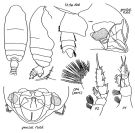 issued from : E.L. Markhaseva in Proc. Zool. Inst. RAN, St. Petersburg, 1996, 268. [p.263, Fig.209]. Female (from NW Pacif.: Kuril Trench). Nota: Cephalothorax about 3.5-4.0 times longer than urosome. Posterior corners of last thoracic segment rounded, symmetrical. Genital segment symmetrical, nearly as long as wide, and nearly as long as 2 following segments together; ventral swelling strongly protruded, widest in its posterior part (lateral view). Urosomal segments 1-3 with hairs on the surface. Body surface of some specimens with chitinous thickenings. A1 longer than cephalothorax reaching urosomal segments 2-3. Exopodite of P1 3-segmented. Endopodite of P2 indistinctly 2-segmented (in Sars, 1925) or distinctly segmented (in A. Scott, 1909). Coxopodite of P4 with about 30 thin fan-like spines (Sars, 1925 noted 14 spines).
|
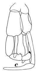 Issued from : W. Vervoort in Zool. Verh., Leiden, 1949, 5. [p.47, Fig.21,c]. As Pseudochirella obtusa. Male (from Banda Sea): c, P5.
|
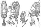 Issued from : G.O. Sars in Résult. Camp. Scient. Prince Albert I, 69, pls.1-127 (1924). [Pl.XXVI, figs.10-15]. Female (near Azores Is.): 10, habitus (dorsal); 11, idem (lateral left side); 12, forehead (lateral); 13, basipodal segment of P4. Male: 14, habitus (lateral left side); 15, P5. Head and 1st thoracic segment with suture more or less visible Nota: Strong development of setae on A1. Nota Male after Markhaseva (1996, p.263): Cephalon and 1st thoracic segment indistinctly separated. Posterior corners of last thoracic segment rounded, without spines. Cephalothorax about 3 times longer than urosome; Exopodal segment 2 of left P5 with 3 teeth in distal part of segment.
|
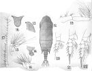 issued from : A. Scott in Siboga-Expedition, 1909, XIX a. [Plate XIII, Figs.1-12]. As Euchirella granulata. Female (from Manipa Strait): 1, habitus (dorsal); 2, forehead (lateral); 3, last thoracic and genital segments (left side); 4, rostrum; 5, portion of the integument with fine granulations (highly magnofied); 6, A1; 7, A2; 8, Mx1; 9, P1; 10, P2; 11, P4 (basal portion only).
|
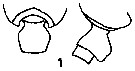 Issued from : E.L. Markhaseva in USSR Acad. Sci., Zool. Inst., Explor. Fauna Seas. Marine Plankton, 1989, 41 (49). p.52, Fig.1]. Female: Thoracic segment 5 and genital segment. Nota from the key: 1 - Genital segment symmetrical.
2 - Thoracic posterior corners symmetrical.
3 - Exopodite 2 of A2 with 3 setae. 4 - Thoracic segments 4 and 5 separate. 5 - Genital segment with ventral protuberance backward. Genital segment (in lateral in lateral view) nearly 2 times large than the following segment. 6 - Body length: 4.8-6,1 mm.
|
 Pseudochirella dubia Pseudochirella dubia Male: 1 - Exopdite 2 of left P5 with 3 teeth in distal part of segment. 2 - Posterior corners of last thoracic segment without spines.
| | | | | Compl. Ref.: | | | Jespersen, 1934 (p.67); Sewell, 1948 (p.500); Grice & Hulsemann, 1967 (p.15); Razouls & al., 2000 (p.343, Appendix); Park & Ferrari, 2009 (p.143, Table 4, Appendix 1) | | | | NZ: | 9 | | |
|
Distribution map of Pseudochirella dubia by geographical zones
|
| | | | | | | | | 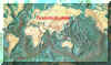 Chart of 1996 Chart of 1996 | |
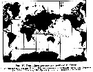 Issued from : E.L. Markhaseva in Issled. Fauny Morei, 1989, 41 (49). [p.55, Fig.10]. Issued from : E.L. Markhaseva in Issled. Fauny Morei, 1989, 41 (49). [p.55, Fig.10].
Geographical distribution of P. dubia.
1: from literature; 2: present occurences. |
| | | | Loc: | | | Antarct. (SW Atlant., Indian), N Atlant. (Azores), Davis Strait, Faroe Is., W Indian, Indonesia-Malaysia, Philippines, Japan Trench, Kuril Is., S Chile.
Type locality: 36°17'N, 28°53'W | | | | N: | 9 | | | | Lg.: | | | (1) F: 4,8; M: ± 4; (5) F: 5,5; (37) F: 6,1-4,8; {F: 4,80-6,10; M: 4,00} | | | | Rem.: | bathy-abyssopelagic.
Sampling depth (Antarct.) : 7000 m. | | | Last update : 05/02/2015 | |
|
|
 Any use of this site for a publication will be mentioned with the following reference : Any use of this site for a publication will be mentioned with the following reference :
Razouls C., Desreumaux N., Kouwenberg J. and de Bovée F., 2005-2026. - Biodiversity of Marine Planktonic Copepods (morphology, geographical distribution and biological data). Sorbonne University, CNRS. Available at http://copepodes.obs-banyuls.fr/en [Accessed February 27, 2026] © copyright 2005-2026 Sorbonne University, CNRS
|
|
|
|




;)
;)
;)
;)
;)
;)

;)
;)