|
|
 |
|
Calanoida ( Order ) |
|
|
|
Spinocalanoidea ( Superfamily ) |
|
|
|
Spinocalanidae ( Family ) |
|
|
|
Methanocalanus ( Genus ) |
|
|
| |
Methanocalanus gabonicus Ivanenko, Defaye & Cuoc, 2007 (F,M) | |
| | | | | | | Ref.: | | | Ivanenko & al., 2007 (p.40, Descr.F,M, figs.F,M, Rem.: internal morphology) | 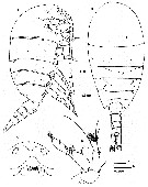 issued from : V.N. Ivanenko, D. Defaye & C. Cuoc in Cah. Biol. Mar., 2007, 48. [p.41, Fig.1]. Female (from off Gabon): A-B, habitus (lateral and dorsal, respectively); C, forhead (ventral); D, A2. Nota: A1 24-segmented, extending to pedigerous segment 4. A2 endopod shorter than exopodLabrum and paragnaths forming a sort of robust oral cone protruding ventrally.
|
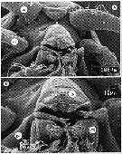 issued from : V.N. Ivanenko, D. Defaye & C. Cuoc in Cah. Biol. Mar., 2007, 48. [p.42, Fig.2, A-B]. Female (SEM photos): A, anterior part of prosome (ventral); B, labrum paragnaths and mandibular gnathobases. Abbreviations: a1 = A1; a2 = A2; lb = labrum; pg = paragnath; gn = gnathobase of mandible. Nota: Labrum oroduced ventrally, distal part with a small row of small setules. Paragnaths: a pair of lobes on pedestal produced ventrally and ornamented with numerous setules.
|
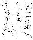 issued from : V.N. Ivanenko, D. Defaye & C. Cuoc in Cah. Biol. Mar., 2007, 48. [p.43, Fig.3]. Female: A, urosome (dorsal); B, A1 (segments 1-17); C, A1 (segments 18-24); D, Md (mandibular palp); E, Md (gnathobase). Nota: Gnathobase of Md elongate, masticatory edge with 8 teeth and 1 seta ( 3 strong teeth separated from 5 smaller teeth by a groove ornamented with setules); exopod 5-segmented, endopod 2-segmented.
|
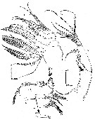 issued from : V.N. Ivanenko, D. Defaye & C. Cuoc in Cah. Biol. Mar., 2007, 48. [p.44, Fig.4]. Female: A, Mx1; A1, proximal basal endite of Mx1; B, Mx2; C, Mxp. Nota: Praecoxal Mx1 with 13 spines; coxal epipodite and coxal endite with 9 and 4 setae, respectively; 1st and 2nd basal endites each with 4 setae; endopod indistinctly 3-segmented bearing 2, 3 and 3 setae, distal segment with row of setules; exopod as a lobe fused to basis and bearing 8 unequal setae and row of setules.
|
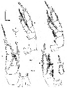 issued from : V.N. Ivanenko, D. Defaye & C. Cuoc in Cah. Biol. Mar., 2007, 48. [p.45, Fig.5]. Female: A, P1 (anterior); B, P2 (anterior); C, distal segment of exopod of P2 (anterior); D, P3 (anterior); F, inner coxal seta of P4 (anterior).
|
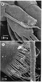 issued from : V.N. Ivanenko, D. Defaye & C. Cuoc in Cah. Biol. Mar., 2007, 48. [p.42, Fig.2, E, F]. Female (SEM photos): E, endopod of left P1 (anterior); F, lobe of endopod of right P1 (anterior view) (= Von Vaupel Klein's organ).
|
 issued from : V.N. Ivanenko, D. Defaye & C. Cuoc in Cah. Biol. Mar., 2007, 48. [p.42, Fig.2, D]. Female (SEM photos): D, genital double-somite (ventral). Nota: genital operculum covered the genital atrrium, and laterallythe atrila slits.
|
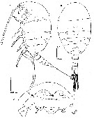 issued from : V.N. Ivanenko, D. Defaye & C. Cuoc in Cah. Biol. Mar., 2007, 48. [p.46, Fig.6]. Male: A-B, habitus (lateral and dorsal, respectively); C, anterior part of cephalosome (ventral). Abbreviations: a1 = A1; a2 = A2; gn = gnathobase of mandible; pm = mandibular palp; lm = labrum. Nota: labrum flat, not produced ventrally, with pair of distolateral bifid extension. Paragnaths absent.
|
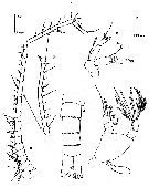 issued from : V.N. Ivanenko, D. Defaye & C. Cuoc in Cah. Biol. Mar., 2007, 48. [p.48, Fig.8]. Male: A, urosome (ventral); B, left A1 (segments 1-18); B, idem (segments 19-23) D, part of right A1 (two-sided arrows indicate homologous segments of right and left A1; E, A2; F, Md (mandibular palp). Nota: A1 asymmetrical; left A1 23-segmented; right A1 22-segmented (differs from left antennule in fusion of segments 19 and 20, shape of segments 13-18). Md gnathobase stylet-like, thin and tubiform, pointed and separated terminally; basis with 2 setae, 1st endopod unarmed, 2nd with 8 setae and row of minute spinules; exopod 5-segmented bearing 1, 1, 1, 1, and 2 setae.
|
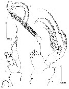 issued from : V.N. Ivanenko, D. Defaye & C. Cuoc in Cah. Biol. Mar., 2007, 48. [p.49, Fig.9]. Male: A, Mx1; B, setae of coxal epipodite of Mx1; C, P1 (anterior).
|
 issued from : V.N. Ivanenko, D. Defaye & C. Cuoc in Cah. Biol. Mar., 2007, 48. [p.49, Fig.9]. Male: A, Mx1; B, setae of coxal epipodite of Mx1; C, P1 (anterior).
|
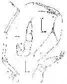 issued from : V.N. Ivanenko, D. Defaye & C. Cuoc in Cah. Biol. Mar., 2007, 48. [p.51, Fig.10]. Male: A, Mx2; B, Mxp; C, P5 (posterior). Copepodd V male: D, P5. Nota: Armature of appendages except P5, similar with that of female.
| | | | | NZ: | 1 | | |
|
Distribution map of Methanocalanus gabonicus by geographical zones
|
| | | | Loc: | | | SE Atlant. (Gabon: continental margin) | | | | N: | 1 | | | | Lg.: | | | (974) F: 1,56; M: 1,57; {F: 1,56; M: 1,57} | | | | Rem.: | Sampling depth: 3151 m.
According to Ivanenko & al. (2007, p.52), in all males, after examination of internal anatomy (semi-thin sections in the prosome), it was not possible to identify any structure corresponding to the digestive tract. These preliminary observations suggest that the male is lacking any digestive system, as supposed from the morphology of the buccal appendages (reductions and modifications of labrum, paragnaths, mandible, maxillule, maxilla and maxilliped).
The feeding apparatus of the adult females, as well as the male copepodid V is characterized by the protruding oral cone formed by the well developed labrum and paragnaths, the elongate mandibular gnathobase with wide masticatory blade armed with stout teeth, the stout setae on the praecoxal arthrite of the maxillule and the endites of the maxilla, the small number of setae on the proximal segments of maxilliped. All these morphological features suggest that the females probably feed on small particles such as detritus and bacterial flocks, grasping from or near the bottom environment. The lower abundance of males than females and the features of their mouthparts suggest that males do not feed and die soon after copulation.
The individuals were found in hydrocarbon fluids, as few centimetres above vesicomyid and mytilid bivalves aggregating at the giant pockmark Regab on the Gabon continental margin, swarming in deep sea chemosynthetic environment. | | | Last update : 17/01/2015 | |
|
|
 Any use of this site for a publication will be mentioned with the following reference : Any use of this site for a publication will be mentioned with the following reference :
Razouls C., Desreumaux N., Kouwenberg J. and de Bovée F., 2005-2026. - Biodiversity of Marine Planktonic Copepods (morphology, geographical distribution and biological data). Sorbonne University, CNRS. Available at http://copepodes.obs-banyuls.fr/en [Accessed February 28, 2026] © copyright 2005-2026 Sorbonne University, CNRS
|
|
 |
 |


