|
|
 |
|
Monstrilloida ( Order ) |
|
|
|
Monstrillidae ( Family ) |
|
|
|
Monstrillopsis ( Genus ) |
|
|
| |
Monstrillopsis chilensis Suarez-Morales, Bello-Smith & Palma, 2006 (F,M) | |
| | | | | | | Ref.: | | | Suarez-Morales & al., 2006 (p.97, Descr.F, figs.F); Suarez-Morales & al., 2008 (p.221, figs.F,M); Suarez-Morales, 2011 (p.3, fig.F) | 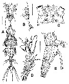 Issued from : E. Suarez-Morales, F.C. Ramirez & C. Derisio in Contr. Zool., 2008, 77 (4). [p.221, Fig.4]. Female (from 54°53'S, 67°29'W): A, habitus (dorsal); B, cephalic area (lateral), showing cuticular processes and ornamentation; C, same (ventral); D, left A1 (dorsal); E, urosome (lateral). Scale bars: 300 µm (A); 100 µm otherwise.
|
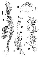 Issued from : E. Suarez-Morales, F.C. Ramirez & C. Derisio in Contr. Zool., 2008, 77 (4). [p.221, Fig.3, Left panel]. Male: A, habitus (lateral); B, cephalic area (ventral), showing cuticular ornamentation; C, same (lateral); D, right A1 (dorsal); E, urosome (lateral), showing genital lappets. Scale bars: 100 µm (A); 50 µm otherwise.
|
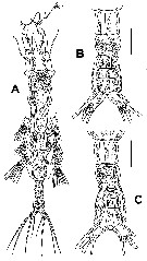 Issued from : E. Suarez-Morales, F.C. Ramirez & C. Derisio in Contr. Zool., 2008, 77 (4). [p.221, Fig.3, Right panel]. Male: A, habitus (dorsal); B, urosome (dorsal); C, same (ventral). Scale bars: 100 µm (A); 50 Mm otherwise.
|
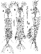 Issued from : E. Suarez-Morales, A. Bello-Smithj & S. Palma in Zool. Anz., 2006, 245. [p.98, Fig.2]. Female (from coastal area off Valparaiso): A, habitus (ventral); B, habitus (dorsal); C, habitus (lateral); D, terminal part of right ovigerous spine. Nota: - Cephalothorax representing 52.3% of total body length. - Oral papilla slightly protuberant, located anteriorly, less than 15% of way back along ventral surface of cephalothorax. - Urosome consisting of 4 somites: 25th pedigerous somite bearing P5, genital double-somite, free postgenital somite, and free anal somite bearing pair of caudal rami. - 5th pedigerous somite with anterior half laterally produced to form pair of rounded processes (this somite representing about 9 % of total body length, slightly longer than 2 postgenital somites together); somite with deep corrugations on anterior outer margins (fig. 4 F). - Ovigerous spines relatively long, equal to about 66% of total body length and about 1.27 times as long as cephalothorax; left spine broken distally in holotype, unbroken right spine distally attenuate. Adhering egg cluster (with 4 rounded eggs measuring 0.40-0.047 mm in diameter) on proximal section of spines, all covered by thin, gelatinous sheath. - Caudal rami subrectangular, widely divergent, approximately 2.1 times longer than wide, each ramus bearing 4 well-developed setae: 2 terminal, 1 outer and 1 on inner margin; innermost seta shortest (0.2 mm), remaining 3 setae noticeable longer, outermost (0.48 mm), outer middle (0.55 mm) and inner middle (0.61 mm).
|
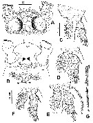 Issued from : E. Suarez-Morales, A. Bello-Smithj & S. Palma in Zool. Anz., 2006, 245. [p.99, Fig.3]. Female: A, cephalic area showing position of sensilla and pigmentation of ocelli (dorsal view); B, cephalic area showing two pairs of ventral cuticular processes (both arrowed); C, basis and rami of P1 (anterior view); D, P2; E, P3 with strongly developed basipodal seta; F, P4. All swimming setae cut short in C-F; G: ornamentation of terminal spiniform seta of P2. Nota: - Cephalic region with rounded forehead bearing pair of well developed sensilla and short ventral cuticular process in rostral position. Dorsal surface of cephalic area with faint pattern of longitudinal cuticular wrinkles arranged in loose pattern, visible only at high magnification (100 x). Anterior ventral surface of cephalic area with pair of subconical processes and field transverse wrinkles between A1 bases and oral paopilla (large arrows in fig. 3B), also visible in lateral view (arrowed in fig.4A). Usual pair of nipple-like processes posterior to subconical processes, radially ridged and furrowed, with postero-medially directed furrows ending in pair of closely set secondary processes (small arrows in fig.3B). - Pair of ocelli (lateral cups of naupliar eye) present, well developed: pigment cups separated by about 3/4 of diameter of one ocellus. Ocelli strongly pigmented medially.
|
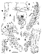 Issued from : E. Suarez-Morales, A. Bello-Smithj & S. Palma in Zool. Anz., 2006, 245. [p.100, Fig.4]. Female: A, vental surface of cephalic region (lateral view) showing cuticular ornamentation, arrowhead pointing to ventral processes; B, 5th pedigerous somite and genital double-somite showing P5 and base of ovigerous spines; (ventral); C, right A1 (dorsal view); D, left A1 (dorsal view), arrowhead pointing ton strongly developed seta ''5''; E, 5th pedigerous somite and genital double-somite showing P5 and base of ovigerous spines (lateral); F, right margin of 5th pedigerous somite in dorsal view showing corrugation of outer proximal margin (arrowed). Nota: - Genital double-somite relatively long, ratio of its length and lengths of two succeeding somites 55 : 20 : 25 = 100. Partial intersegmental division on lateral surface, marked by notch on lateral margin visible in dorsal view. Anterior half of genital double-somite with outer margins expanded laterally, expansions rounded, posterior half unexpanded, with transverse cuticular wrinkles visible in dorsal and lateral view. Insertion point of paired ovigerous spines on proximal half of ventral side of double-somite, spines arising separately from protuberant base visible in lateral view. - P5 medially conjoined at base, unsegmented, each consisting of relatively large outer (exopodal) lobe and inner (endopodal), digitiform lobe. Endopodal lobe relatively short, reaching at most midlength of exopodal lobe, unarmed. Exopodal lobe bearing 3 setae, all of different lengths; innermost seta shortest, middle seta longest, about 5 times longer than inner one, outermost seta about 1.5 times longer than innermost; Innermost seta noticeably thinner than middle and outer setae. All three setae biserially but sparsely setulated. - A1equal to 17.7 % of total body length and 34 % of cephalothorax length. A1 4-segmented, armed with 0-I; I-V; 23-I; 10-VII setae (Arabic numerals) and spines (or spiniform setae) (Roman numerals), plus 2 aesthetascs. In terms of the pattern described by Grygier & Ohtsuka (1995) for female monstilloid antennular armature.
|
 Issued from : E. Suarez-Morales, A. Bello-Smithj & S. Palma in Zool. Anz., 2006, 245. [p.97]. Female: Armature formula of swimming legs P1 to P4.
| | | | | Compl. Ref.: | | | Suarez-Morales, 2011 (p.12) | | | | NZ: | 2 | | |
|
Distribution map of Monstrillopsis chilensis by geographical zones
|
| | | | | | | Loc: | | | SE Pacif. (Chile: off Valparaiso), SW Atlant. (Bahia Brown, Beagle Channel)
Type locality: 71°38.34' S, 33°01.11' W. | | | | N: | 2 | | | | Lg.: | | | (978) F: 1,76 [without furca]; (1029) M: 0,78; {F: 1,76; M: 0,78} | | | | Rem.: | coastal (bottom: 25 m).
After Lee J. & al. (2016, p.421), male genitalia with a relatively short median shaft with its posterolateral corners extended as elongate lappets belong subgroup ''Type II'' (Suarez-Morales & McKinnon, 2014)
For Suarez-Morales & al. (2006, p.101) this species differs from Scott's (1904) M. dubia, the latter species has relatively longer A1 (32.5 % of total body length) than the new species (less than 18 %); both, however, share a relatively long terminal antennular segment. M. dubia (sensu Scott, 1904) has a short, robust genital double-somite (Scott 1904: pl. XIV, Fig.18) 1.2 times wider than; this somite represents about 4 % of the total body length and about 42 % of the urosomal length, whereas the corresponding structure in the bnew species has a different shape and greater relative length with respect to both total body length (9 %) and urosomal length (55 %) Also, the 5th pedigerous somite is relatively smaller in M. dubia (sensu Scott, 194) than in M. chilensis. The anal somite is noticeably longer than the preanal somite in M. dubia and represents 30 % of the urosomal length, whereas in the new species the anal somite is only slightly longer than the preanal somite.
Suarez-Morales & al. (2006, p.103, Table 1): See Morphological characters for comparative morphology of the species assigned to the genus Monstrillopsis by different authorities : M. dubia, M. dubioides, M. fosshageni, M. reticulata, M. sarsi, Cymbasoma gracilis, M. ferrari, M. zernovi, M. angustipes, Monstrillopsis sp.
Key for the identification of the currently known species of Monstrillopsis (female only) after Suarez-Morales & al. (2006, p.105):
1 - Outer lobe of P5 without distal elongation; last antennular segment less than the 45% of A1 length ; body length less than 3 mm...... 2.
2 - Anterior half of 5th pedigerous soůmite laterally protuberant; lateral margins of genital double-somite expanded proximally; A1 representing less than 20% of total body length; 4 caudal setae...... 3.
3 - Middle exopodal seta of P5 longest, inner seta small, about 1/5 of length of middle seta; dorsal cephalic surface smooth; urosomal somites smooth; body less than 2 mm in length. | | | Last update : 07/03/2020 | |
|
|
 Any use of this site for a publication will be mentioned with the following reference : Any use of this site for a publication will be mentioned with the following reference :
Razouls C., Desreumaux N., Kouwenberg J. and de Bovée F., 2005-2026. - Biodiversity of Marine Planktonic Copepods (morphology, geographical distribution and biological data). Sorbonne University, CNRS. Available at http://copepodes.obs-banyuls.fr/en [Accessed February 07, 2026] © copyright 2005-2026 Sorbonne University, CNRS
|
|
 |
 |


