|
|
 |
|
Cyclopoida ( Order ) |
|
|
|
Oncaeidae ( Family ) |
|
|
|
Epicalymma ( Genus ) |
|
|
| |
Epicalymma bulbosa Böttger-Schnack, 2009 (F,M) | |
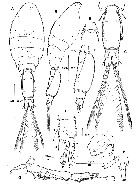
| | | | | | | Ref.: | | | Böttger-Schnack, 2009 (p.1029, figs.F,M); Böttger-Schnack & Schnack, 2013 (p.4: Table 1) |  Issued from R. Böttger-Schnack in J. Plankton Res., 2009, 31 (9). [p.1031, Fig.1]. Female (from Red Sea): A-B, habitus (dorsal and lateral, respectively); C, posterior part of P4-bearing somite and urosome (dorsal); D, urosome (lateral); E, P5-bearing somite and anterior part of genital double-somite (ventral); F, caudal ramus (dorsal, individual elements numbered using Roman numerals); G, A1; H, P5 (lateral); I, P6, armature not clearly discerned (stippled). Nota: Prosome 2.3 times length of urosome, excluding caudal rami, 1.9 times urosome length including caudal rami. A1 6-segmented; relative lengths (%) of segments measured along posterior non-setiferous margin 7.7 : 17.5 : 44.3 : 11.4 : 4.7 : 14.4. Segment 1 ornamented with short row of spinules. Proportional lengths (%) of urosomites 8.8 : 67.1 : 4.9 : 4.6 : 14.6 and proportional lengths (%) of urosomites and caudal rami 7.5 : 57.5 : 4.1 : 4.0 : 12.5 : 14.4. Genital double-somite elongate, 2.1 times as long as maximum width (measured in dorsal aspect) and 2.8 times as long as postgenital somites combined; lateral margins almost straight, largest width measured along midregion from 1/4 distance of posterior margin. Paired genital apertures located dorsally at about 1/4 distance from anterior margin of genital double-somite; armature hardly discernible, possibly 1 or 2 diminutive spinules (stippled in fig.1 i). Anal somite 1.8 times wider than long; slightly shorter than caudal rami. Dorsal surface with paired dorsal sensillae anterior to anal opening, pair of secretory pores at midregion of somite and patch of small middorsal spinules near posterior margin. Posterior margin of somite finely serrate ventrally and laterally. Caudal ramus about 1.3 times as long as wide measured along outer margin, with conspicuous expansion on dorsal surface surrounding insertion of seta VII. Armature consisting of 6 elements; seta II and III small and slender, spiniform and unornamented; seta IV and V very long and resilient, almost equal in length, bulbous ventricle on the inner cavity of both setae absent; seta IV displaced to midregion of distal margin and slightly bent near base, ornamented with short spinules along outer margin and long, fine pinnules along inner margin; seta V with outer margin unornamented, inner margin ornamented with few short spinules anteriorly and long, fine pinnules at posterior part; seta VI about same length as seta III, spiriform and unnipinnate; seta VII unornamented and very long, almost as long as seta IV and V, biarticulation at base not discernible, probably absent. Inner margin of caudal ramus unornamented. Dorsal anterior surface with minute secretory pore nerar insertion of seta II (fig.1F). P5 (fig. 1H) : comprised of short outer basal seta, reaching almost as far as genital apertures and being sparsely plumose, and small exopod segment delimited from somite. Exopod about as long as wide, ornamented with a small spinous process laterally and armed with single very long, plumose seta,0 reaching about 4/5 length of genital double-somite, almost as far as paired pore on dorsal surface. P6 represented by operculum closing off each genital aperture; armature not clearly discernible, possibly represented by 1-2 minute spinules (stippled in fig.1I)
|
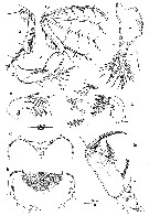
 Issued from R. Böttger-Schnack in J. Plankton Res., 2009, 31 (9). [p.1032, Fig.2]. Female: A, A2 (anterior, lateralelements numbered using Roman numerals, distal elements numbered using capital letters [a, distal endopod segment, posterior, setae A-D omitted]; B, labrum (anterior); C, same (posterior); D, Md (showing individual elements numbered using capital letters); E, Mx1; F, Mx2 (seta on inner margin of allobasis figured separately); G, Mxp (posterior; arrow indicating long, basally swollen spinule on syncoxa). Nota: A2 3-segmented. Coxobasis with row of long spinules along outer margin and few long, fine spinules at inner margin; posterior surface with few additional short denticles; with long bipinnate seta at inner distal corner. Endopod segments unequal in length, distal segment about 1.5 times longer than proximal one. Labrum bilobed. Distal (ventral) margin of each lobe with 1 strong marginal tooth medially, long row of small spinules or denticles at outer ventral margin and row of 7-8 large, strong denticles alonginner margin, all ornamentation elements inserting posteriorly. Median concavity covered anteriorly by several overlapping rows of hyaline spatulated setules. Anterior surface (fig.2B) unornamented, except for large secretory pore posterior to weakly developed median bulge. Posterior wall of medial cocavity with 1 large chirinized spinous tooth, showing 4-fold upper edge (fig. 2C). Ornamentation on posterior surface not discerned. Md gnathobase with 5 elements: 3 setae and 2 blades. Ventral element (A) slightly shorter than ventral blade (B), with long fine setules along dorsal side; ventral blade strong and spiniform, unornamented; dorsal blade (C) strong and broad, spinulose along dorsal margin; seta D almost as long as blades B and C, spinulose along dorsal margin; dorsal element (E) longest, setiform and bipinnate. Mx1 weakly bilobed, no surface unornamention discernible. Inner lobe (praecoxal arthrite) with 3 elements: outermost element spiniform, slightly swollen at base, ornamented with spinules unilaterally and 2 coarse spinules, middle element setiform and unipinnate; innermost element located along concave inner margin close to other elements, sparsely bipinnate. Outer lobe with 4 setiform elements, which are bare or unipinnate, element next to innermost seta longest and swollen at base, outermost element shortest. Mx2 2-segmented, allobasis slightly longer than syncoxa. Syncoxa unarmed, surface ornamented with cluster of long spinules and 1 large secretory pore. Allobasis produced distally into slightly curved claw bearing 2 rows of sgtrong spinules along medial margin; outer margin with strong seta not extending to tip of allobasal claw; ornamented with long spinules bilaterally, tubular extension on tip of seta not discernible; inner margin with long naked seta (figured separately in fig. 2F) and strong basally swollen spine with double row of long fine spinules along the medial margin and 2 spinules along outer margin. Mxp 4-segmented (syncoxa, basis and 2-segmented endopod). Syncoxa unarmed, posterior surface ornamented with small denticles and 1 long, basally swollen spinule at inner margin (arrowed in fig.2G).
The specific species name is derived from the Latin ''bulbous'', meaning bulbous, and refers to the form of the long, basally swollen spinule on the syncoxa of the female maxilliped.
|
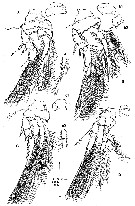
 Issued from R. Böttger-Schnack in J. Plankton Res., 2009, 31 (9). [p.1034, Fig.3]. Female: A, P1 (posterior; intercoxal sclerite figured separately, tubular extensions on outer exopodal spines exemplarily enlarged on exopod-1 and -3 [a, terminal part of endopod, anterior, showing apical processes]; B, P2 (anterior; b1, intercoxazl scerite, posterior, slightly damaged; b2, inner basal margin, posterior, arrow indicating pore); C, P3 (anterior; c1, intercoxal sclerite, anterior; c2, endopod-2 and proximal part of endopod-3, anterior, showing spinular row indicating region of fusion of segments); D, P4 (anterior; intercoxal sclerite figured separately).
|
 Issued from R. Böttger-Schnack in J. Plankton Res., 2009, 31 (9). [p.1035, Table II]. Armature of swimming legs P1 to P4.
|
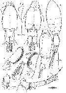
 Issued from R. Böttger-Schnack in J. Plankton Res., 2009, 31 (9). [p.1036, Fig.4]. Male: A, habitus (dorsal; ornamentation of caudal setae not shown); B, A1 (length of distal aesthetasc not fully discerned); C, Mxp (anterior; small spine basally fused to inner proximal corner of claw figured separately [c1, same, posterior; c2, maxillipedal basis, medial view, claw omitted, arrow indicating seta within longitudinal cleft]); D, urosome (dorsal; caudal ramus setae IV, V and VII not fully shown); E, same (ventral); F, urosome (lateral); G, P5 (dorsolateral); H, caudal ramus (dorsal; showing dorsal expansion, setae IV, V and VII omitted). Nota: Proportional lengths (%) of urosomites (excluding caudal rami) 10.4 : 63.2 : 4.1 : 4.7 : 5.2 : 12.4. Proportional lengths (%) of urosomites (caudal rami included) 8.8 : 53.5 : 3.5 : 3.9 : 4.4 : 10.5 : 15.4. Anal somite with length to width as in female, pore pattern as in female. A1 4-segmented; distal segment corresponding to fused segments 4-6 of female. Segment 1 ornamented with short row of spinules as in female. P5 exopod not delimited from somite, located latero-ventrally, armature as in female; outer basal seta as in female. P6 represented by posterolateral flap closing off genital aperture on either side, unornamented; posterolateral corners not protruding laterally and not well discernible in dorsal aspect. Spermatophore oval of variable size according to state of maturity; swelling of spermatophore during development not affecting shape and relative size of genital somite (fig. 4 D, F)
|
 Issued from R. Böttger-Schnack in J. Plankton Res., 2009, 31 (9). [p.1037, Fig.5]. Male: A, P1 (anterior); B, P2 endopod (anterior; inner setae omitted). Nota: P1-P4 with armature and ornamentation as in female.
| | | | | NZ: | 1 | | |
|
Distribution map of Epicalymma bulbosa by geographical zones
|
| | | | Loc: | | | Red Sea (N-S), Gulf of Aqaba
Type locality: 27°25.00'N, 34°04.96'E. | | | | N: | 1 | | | | Lg.: | | | (1032) F: 0,302-0,347; M: 0,292-0,294; {F: 0,302-0,347; M: 0,292-0,294} | | | | Rem.: | For Böettger-Schnack (2009, p.1038) this species is closely related to E. vervoorti. Females exhibited some variability in the length-to-width ratio of the genital double-somite, with the typical form exhibiting a smaller length-to-width ratio (1.8:1) than the elongate form (2.3:1). | | | Last update : 02/12/2016 | |
|
|
 Any use of this site for a publication will be mentioned with the following reference : Any use of this site for a publication will be mentioned with the following reference :
Razouls C., Desreumaux N., Kouwenberg J. and de Bovée F., 2005-2026. - Biodiversity of Marine Planktonic Copepods (morphology, geographical distribution and biological data). Sorbonne University, CNRS. Available at http://copepodes.obs-banyuls.fr/en [Accessed January 31, 2026] © copyright 2005-2026 Sorbonne University, CNRS
|
|
 |
 |


