|
|
 |
|
Monstrilloida ( Order ) |
|
|
|
Monstrillidae ( Family ) |
|
|
|
Maemonstrilla ( Genus ) |
|
|
| |
Maemonstrilla polka Grygier & Ohtsuka, 2008 (F) | |
| | | | | | | Ref.: | | | Grygier & Ohtsuka, 2008 (p.470, Descr. F, figs.F, Rem.) | 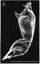 issued from : M.J. Grygier & S. Ohtsuka in Zool. J. Linnean Soc., 2008, 152. [p.471, Fig.7, A]. Female (paratype from Ishigaki Island): A, habitus (lateral; some legs removed). Scale bar = 1.0 mm. Nota: - Spherical spots of red pigment throught body. - Cuticular ridges of cephalothorax sporadically spinulose but lacking side branches, discontinuous in anterior ventrolateral region; reticulation uniform in size, enclosing clusters of small, cuneiform cuticular figures. - Posterior dorsal margin of genital compound somite and penultimate segment each slightly produced into 6 flanges bearing minute spinules of same size and density as those over rest of dorsum. Posteroventral part of genital compound somite produced into apron-like flap.
|
 issued from : M.J. Grygier & S. Ohtsuka in Zool. J. Linnean Soc., 2008, 152. [p.471, Fig.7, B, C]. Female: B, oral papilla (o) and scars (arrow) (left lateral view); C, detail of scars. Scale bars = 0.100 mm (B); 0.010 mm (C).
|
 issued from : M.J. Grygier & S. Ohtsuka in Zool. J. Linnean Soc., 2008, 152. [p.472, Fig.8]. Female: right A1 (ventral view) (setules omitted, setal designation after Grygier & Ohtsuka, 1995, fig.6). Scale bar = 0.100 mm. Nota: Spiniform 2v-setae and 3-seta on A1 about twice as long as 4-setae; apical 6-setae minutely spinulose.
|
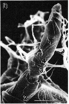 issued from : M.J. Grygier & S. Ohtsuka in Zool. J. Linnean Soc., 2008, 152. [p.471, Fig.7, D]. Female: D, left A1 (outer view), foreshortened due to viewing angle, more distal segments numbered (2-4), arrow indicating branches of b5-seta. Scale bar = 0.100 mm.
|
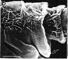 issued from : M.J. Grygier & S. Ohtsuka in Zool. J. Linnean Soc., 2008, 152. [p.473, Fig.9, D]. SEM. Female: D, genital compound somite and penultimate segment of urosome (lateral view), anterior to left, showing apron-like posteroventral protrusion of compound somitre (arrow). Scale bar = 0.050 mm.
|
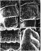 issued from : M.J. Grygier & S. Ohtsuka in Zool. J. Linnean Soc., 2008, 152. [p.474, Fig.10]. SEM. Female: A, dorsal surface of posterior part of cephalothorax and free pedigers 1 and 2, anterior at top; B, dorsal surface of free pedigers 3 and 4, anterior at top; C, dorsal surface of penultimate segment and telson (t) of urosome, anterior at top; D, proximal part of coxa of P1, showing spinulose outer lobes (arrows), anterior to left. Scale bars = 0.100 mm (A, B); 0.020 mm (C, D).
|
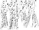 issued from : M.J. Grygier & S. Ohtsuka in Zool. J. Linnean Soc., 2008, 152. [p.475, Fig.11]. Female: A, right P1 (anterior); B, left P2 (posterior); C, outer apical exopodal seta of left P2; D, right P3 (anterior); E, left P4 (posterior). Slide-mount of legs P1 to P4, showing diagnostic red spots and invaginations or vestigial buttons replacing proximal inner endopodal seta of each leg (arrows), setules omitted and some setae cut short. Scale bar = 0.200 mm (A, B, D, E); 0.100 mm in C. Nota: - Outer proximal part of coxa of P1-P4 produced into low, oval lobes armed with spinules slightly larger than those on rest of coxa.
|
 issued from : M.J. Grygier & S. Ohtsuka in Zool. J. Linnean Soc., 2008, 152. [p.473, Fig.9, E]. SEM. Female: E, P5 (arrow). Scale bar = 0.100 mm.
|
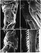 issued from : M.J. Grygier & S. Ohtsuka in Zool. J. Linnean Soc., 2008, 152. [p.476, Fig.12]. SEM. Female: A, basis and 1st exopodal segment of P1, outer view, proximal at top and anterior to right, showing 1 seta on each segment, anterior pore near basis seta (arrow), and spinules patches on exopod; B, 3rd segment of endopod (right) and exopod (left) of P2, anterior view showing distal pore on each segment (arrows); C, proximal endopodal segment of P2, anterior view, showing vestigial button-like seta (arrow); D, detail of outer apical exopodal seta. Scale bars = 0.020 mm (A-C); 0.010 mm in D.
|
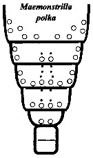 issued from : M.J. Grygier & S. Ohtsuka in Zool. J. Linnean Soc., 2008, 152. [p.499, Fig.29]. Female: Dorsal and lateral pore and pit seta patterns, from rear of cephalothorax through genital compound somite. Symbols: dots (three sizes) = pores; larger circles = pits of pit setae. Pattern based on SEM and light microscopical examination.
|
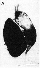 issued from : M.J. Grygier & S. Ohtsuka in Zool. J. Linnean Soc., 2008, 152. [p.477, Fig.13, A]. A, ovigerous female (holotype from Ishigaki Island). photomicrographs of preserved specimen. Scale bar = 0.500 mm. Nota: In lateral view, egg mass appearing bigger than copepod itself (e.g. 2.07 mm long, 1.28 mm high), but width slightly less than half that of cephalothorax (within which all eggs were formerly stored). Mean egg diameter 0.0344 mm (n = 15).
|
 issued from : M.J. Grygier & S. Ohtsuka in Zool. J. Linnean Soc., 2008, 152. [p.462, Fig.1, A, B]. Measurements taken from specimens in species of Maemonstrilla: Lengths in lateral view from example M. polka (A) and in dorsal view (example M. turgida (B).
|
 Issued from : M.J. Grygier & S. Ohtsuka in Zool. J. Linnean Soc., 2008, 152. [p.493]. Key to the Ryukyu species of the Maemonstrilla hyottoko species-Group. M. polka Female: 1 - Cuticle of cephalothorax, A1, lateral sides of trunk, dorsum telson, and caudal rami reticulated; outer faces of P1-P4 and dorsum of free pedigers, genital compound somite, and penultimate somite spinulose. Cephalothoracic reticulations comprising ridges with abundant or sparse spinules; simple or complex cuticular ornamentation within at least some meshes. 2 - Oral papilla large and conical, directed ventrally. Genital compound somite distinctly divided transverse ridge. 3 - P1-P4 each with two low, rounded lobes on outer proximal part of coxa; rest of outer face of coxa with fields of minute spinules separated by bare lanes. Cephalothoracic reticulations lacking side branches but bearing sporadic or sparse spinules; meshes uniformly polygonal over most surface. 4 - Proximal coxal lobes of P1-P4 bearing spinules only slightly larger than those on rest of coxa. Spherical pigmented spots (red when fresh) found throughout body. Posterroventral part of genital compound somite produced into apron-like flap; posterodorsal margins of this somite and penultimate somite not denticulate. Body length > 2mm [after 3 ovigerous females collected].
Compare with M. simplex, M. hyottoko, M. polka, M. spinicoxa.
|
 Issued from : M.J. Grygier & S. Ohtsuka in Zool. J. Linnean Soc., 2008, 152. [p.477]. Maemonstrilla polka Female (from south coast of Ishigaki Island). - Spherical spots of red pigment throughout body. - Cuticular ridges of cephalothorax sporadically spinulose but lacking side branches, discontinuious in anterior ventrolateral region; reticulations uniform in size, enclosing clusters of small, cuneiform cuticular figures. - Spiniform 2v-setae and 3-seta on A1 about twice as long as 4-setae; apical 6-setae minutely spinulose.- Outer proxuimal part of coxa of P1-P4 produced into 2 low, oval lobes armed with spinules slightly larger than those on rest of coxa.
- Posterior dorsal margin of genital compound somite and penultimate segment each slightly produced into 6 flanges bearing minute spinules of same size and density as those over rest of dorsum. Posteroventral part of genital compound somite produced into apron-like flap.
| | | | | NZ: | 1 | | |
|
Distribution map of Maemonstrilla polka by geographical zones
|
| | | | | | | Loc: | | | S Japan (Ishigaki Island) | | | | N: | 1 | | | | Lg.: | | | (1077) F: 2,56-2,66 *
*: sum of lengths of cephalothorax, metasome and urosome. | | | Last update : 23/04/2020 | |
|
|
 Any use of this site for a publication will be mentioned with the following reference : Any use of this site for a publication will be mentioned with the following reference :
Razouls C., Desreumaux N., Kouwenberg J. and de Bovée F., 2005-2026. - Biodiversity of Marine Planktonic Copepods (morphology, geographical distribution and biological data). Sorbonne University, CNRS. Available at http://copepodes.obs-banyuls.fr/en [Accessed February 01, 2026] © copyright 2005-2026 Sorbonne University, CNRS
|
|
 |
 |


