|
|
 |
|
Calanoida ( Order ) |
|
|
|
Clausocalanoidea ( Superfamily ) |
|
|
|
Stephidae ( Family ) |
|
|
|
Stephos ( Genus ) |
|
|
| |
Stephos boettgerschnackae Krsinic, 2012 (F,M) | |
| | | | | | | Ref.: | | | Krsinic, 2012 (p.1527, Descr.F,M, figs.F,M) | 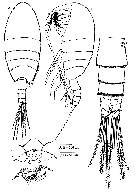 Issued from : F. Krsinic in Crustaceana, 2012, 85 (12-13). [p.1528, Fig.1]. Female (from 14°31'44.3''N, 45°17'11.4''E): A-B, habitus (dorsal and lateral, respectively); C, rostral area (ventral); D, urosome and caudal rami (dorsal). Nota: Cephalon and 1st pediger separate, 4th and 5th fused. Rostral area with 2 sensilla medially, rostrum absent. Nauplius eye absent. Prosome 2.3 times as long as wide. last pediger segment asymmetrical, longer on right side as a rounded knob. Ratio prosome / urosome (including caudal rami) 2.9 : 1. Urosome 4-segmented; somites with hyaline frill along posterior margins. Proportional lengths of urosomites and caudal rami 37 : 18 : 15.6 : 11 : 18.4 = 100. Caudal rami symmetrical, about as long as wide, inner margin ornamented with fine spinules distally; ramus with 4 terminal setae (corresponding to setae III-VI, seta I absent, setae II and VII small).
|
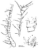 Issued from : F. Krsinic in Crustaceana, 2012, 85 (12-13). [p.1530, Fig.2]. Female: A, genital double-somite (lateral); B, same (ventral); C-D; A1 (segments 1-15 and separated segments 16-24). h.s. = hyaline sheath, gp. = gonopores, op. = operculum, s.r. = seminal receptacle, eg.d. = egg-laying duct. Nota: A1symmetrical, 24-segmented, reaching almost to end of caudal ramiGenital double-somite 1.6 times as long as wide, strongly produced postero-ventrally, slightly asymmetrical in ventral view, tuft of spinules on right anterio-lateral area. External genital area asymmetrical in ventral view, proximally with oval bulge of egg-laying duct, paired gonopores situated laterally on right side near posterior edge, seminal receptacle present on left side only; single operculum produced into spiniform process. Genital area covered by hyaline sheath, proximally reaching to third pedigerous somite on the right side, and ventrally surpassing genital double-somite (this probably belongs to spermatophore as an attachment substance). Spermatophore sac small, reaching to end of caudal rami.
|
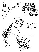 Issued from : F. Krsinic in Crustaceana, 2012, 85 (12-13). [p.1531, Fig.3]. Female: A, A2; B, Md (mandibular palp); C, Md (mandibular gnathobase); D, Mx1; E, Mx2; F, Mxp. Nota: A2 biramous with exopod longer than endopod. Coxa with 1 seta and basis with 2 short setae distally. Exopod 7-segmented, 1.4 times as long as endopod, setal armature 1, 3, 1, 1, 1, 1, 4. Endopod 2-segmented, distal segment armed with 2 transverse rows of setules, 8 unequal setae on lateral margin and 7 setae on apex. Md: gnathobasecutting edge with isolated unicuspid tooth, 8 heterogeneous teeth and 1 dorsal spinulose seta; row of spinules distally on ventral margin and dorsal margin distally and medially.. Palp biramous, basis longer than wide with 4 setae on inner medial margin. Endopod 2-segmented, 1st segment with 4 unequal setae and terminal segment truncate bearing 10 setae. Exopod 5-segmented (setal formula 1, 1, 1, 1, 2, respectively) Mx1: praecoxal arthrite bearing 9 stout marginal spines and 4 elements on posterior surface. Coxal epipodite armed with 9 and coxal endite with 3 setae.Proximal basal endite with 4 setae, distal basal endite with 5, basal exite unarmed. Exopod bearing 11 setae. Endopod articulated to basis, indistintly 3-segmented (setal formula 4, 4, 7, respectively). Mx2: praecoxal endites with 6 and 3 setae, respectively. Coxal endites each with 3 setae. Allobasis with proximal endite powerfully developed with 4 setae, 1 stouter then rest. Endopod 3-segmented. Mxp 7-segmented. Praecoxal endite without seta; syncoxal endites, setal formula 2, 4, 3 on medial margin, distal seta thorn-like; distal margin with row of spinules. Basis with longitudinal row of spinules and with 3 setae, plus 2 on incorporated 1st endopodal segment; free endopod 5-segmented (setal formula 4, 4, 3, 3+1, 4, respectively).
|
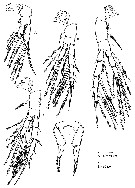 Issued from : F. Krsinic in Crustaceana, 2012, 85 (12-13). [p.1533, Fig.4]. Female: AD, P1 to P4, respectively; E, P5. Nota: P5 symmetrical, uniramous, 3-segmented, proximal segment fused to intercoxal sclerite. 3rd segment longer than segment 2, tapering apically, transverse comb of 2 or 3 spinules at mid-length and armed with spinules distally along inner and outer margin; distal segment armed with single small spine on anterior surface.
|
 Issued from : F. Krsinic in Crustaceana, 2012, 85 (12-13). [p.156, Table I]. Female: Armature formula of the swimming legs P1 to P4. Roman numerals = spines, arabic numerals = setae. Nota: Armature of swimming legs P1 to P4 as that of female.
|
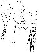 Issued from : F. Krsinic in Crustaceana, 2012, 85 (12-13). [p.1534, Fig.5]. Male: A-B, habitus (dorsal and lateral, respectively); C, urosome (dorsal). Nota: Prosome 2.3 times as long as wide. Posterior prosome asymmetrical, longer on left side. Rostral area as in female. Mouthparts for male as that of female. Prosome /urosome ratio : 2.5 : 1. Urosome of 5 somites. Proportional lengths of urosomites 21 : 24 : 21 : 23 : 11 = 100. Genital somite slightly asymmetrical viewed dorsally, wider than long, genital aperture located on left side. Caudal rami as in female.
|
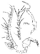 Issued from : F. Krsinic in Crustaceana, 2012, 85 (12-13). [p.1535, Fig.6]. Male: A-B, A1 (segments 1-15 and 17-24); C, left P5; D, right P5. Nota: A1 similar to those of female, 24-segmented with same disposition of setae, but differences in number and position of aesthetascs; segment 1 with 3 strongly enlarged aesthetascs, sement 2 with 4 aesthetascs and segments 3, 7, 17, 20 and 21 with 1 aesthetasc each. Armature of A2, mouthparts and swimming legs 1 to 4 as that of female. P5 uniramous and asymmetrical. Right leg 4-segmented; left leg 5-segmented, segments 1 to 4 unarmed, segment 4 large and strongly expanded, distal end with bulge, outer side regularly rounded, inner margin with outer bulge at midlength; segment 5 complex, elongate and shorter than segment 4, with longitudinal row of 8 short and unequal lamellae on anterior medially surface, first 4 leaf-shaped and others claw-like lamellae; on distal surface 4 lamellae, distal double longer than proximal leaf-like lamellae; on tip 2 long claw-like lamellae.
| | | | | Compl. Ref.: | | | Krsinic, 2015 (p.9: Rem.) | | | | NZ: | 1 | | |
|
Distribution map of Stephos boettgerschnackae by geographical zones
|
| | | | | | | Loc: | | | N Adriatic Sea (near Rijeka, Croatia) | | | | N: | 1 | | | | Lg.: | | | (1100) F: 0.89-0.93; M: 0.82-0.90; {F: 0.89-0.93; M: 0.82-0.90} | | | | Rem.: | anchialine cave.
For Krsinic (2012, p.1536) the species belongs to Type III, where segment 4 of the male left P5 is swollen (see in Genus Stephos, types of classification from Bradford-Grieve, 1999 a, p.25, Table 1) After Krsinic (2015, p.9) the species is widely distributed along the eastern coast of the Adriatic, and is present in almost all anchialine habitats from Rovinj (N Adriaric Sea) to the Island of Hvar (Central Adriatic); the bulk of the population was found in Urinj Cave, near Rijeka (N Adriatic) (see Cukrov & al., 2006 for hydrographic and chemical data). | | | Last update : 11/05/2016 | |
|
|
 Any use of this site for a publication will be mentioned with the following reference : Any use of this site for a publication will be mentioned with the following reference :
Razouls C., Desreumaux N., Kouwenberg J. and de Bovée F., 2005-2026. - Biodiversity of Marine Planktonic Copepods (morphology, geographical distribution and biological data). Sorbonne University, CNRS. Available at http://copepodes.obs-banyuls.fr/en [Accessed February 10, 2026] © copyright 2005-2026 Sorbonne University, CNRS
|
|
 |
 |


