|
|
 |
|
Calanoida ( Order ) |
|
|
|
Pseudocyclopoidea ( Superfamily ) |
|
|
|
Pseudocyclopidae ( Family ) |
|
|
|
Pseudocyclops ( Genus ) |
|
|
| |
Pseudocyclops schminkei Chullasorn, Ferrari & Dahms, 2010 (F,M) | |
| | | | | | | Ref.: | | | Chullasorn & al., 2010 (p.37, Drescr. F,M, figs.F,M); Laakmann & al., 2019 (p.330, fig. 2, 3, phylogenetic relationships) | 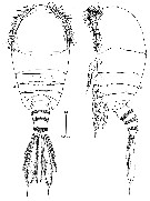 issued from : S. Chullasorn, F.D. Ferrari & H.-U. Dahms in Helgol. Mar. Res., 2010, 64; [p.38, Fig.1]. Female (from aquarium, Zamami Is.): A-B, habitus (dorsal and left lateral, respectively).
|
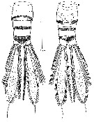 issued from : S. Chullasorn, F.D. Ferrari & H.-U. Dahms in Helgol. Mar. Res., 2010, 64; [p.39, Fig.2]. Female: A-B, urosome (dorsal and lateral, respectively). Nota: Genital double-somite with small point laterally toward anterior edge and ventral ridge posterior to paired, anteroventral oviductal openings; paired copulatory pores anterior to paired, tooth-like, ventral extensions of epicuticule; linear series of indentations from small point to copulatory pore on right side. Anal somite narrow (± telescoped in the preceeding somite). Caudal rami posterior margin extending beyond level of insertion of setae; 1 dorsal, 1 lateral seta and 4 terminal, 3 largest setae with secondary articulation close to their insertion. The 2nd terminal seta from medial seta on the caudal ramus of females is polymorphic; it is usually broad proximally and thin distally on most females, but on a few specimens it may be thick proximally but tapering smoothly distally, like the males. A similar polymorphism has been reported for P. obtusatus asymmetrica by Ummerkutty1 1968 and for P. Xiphophorus by Brugnano & al. (2009).
|
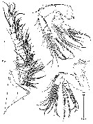 issued from : S. Chullasorn, F.D. Ferrari & H.-U. Dahms in Helgol. Mar. Res., 2010, 64; [p.40, Fig.3]. Female: A, A1; B, A2; C, Md. Nota: A1 17-segmented. A2: endopod 3-segmented; exopod appearing 6-segmented
|
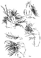 issued from : S. Chullasorn, F.D. Ferrari & H.-U. Dahms in Helgol. Mar. Res., 2010, 64; [p.41, Fig.4]. Female: A, Mx1; B, Mx1 (basis plus rami; C, Mx2; D, endopod of Mx2; E, Mxp. Mx1: Praecoxal endite extending distally, with 5 thick and 3 thin ventral setae, 1 anterior seta and 4 posterior setae; basal exite with 1 seta, proximal endite well-developed with 3 setae, distal endite not extended ventrally with 4 setae; exopod lobate with 4 lateral and 7 terminal setae; endopod 2-segmented, proximal segment with groups of 5 mid-ventral and 4 distoventral setae, distal segment with 6 terminal setae.
|
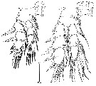 issued from : S. Chullasorn, F.D. Ferrari & H.-U. Dahms in Helgol. Mar. Res., 2010, 64; [p.42, Fig.5]. Female: A, right P1; B, right P2.
|
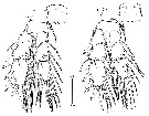 issued from : S. Chullasorn, F.D. Ferrari & H.-U. Dahms in Helgol. Mar. Res., 2010, 64; [p.43, Fig.6, A-B]. Female: A, right P3; B, right P4.
|
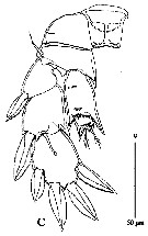 issued from : S. Chullasorn, F.D. Ferrari & H.-U. Dahms in Helgol. Mar. Res., 2010, 64; [p.43, Fig.6, C]. Female: C, right P5. P5: Intercoxal sclerite with complete row of denticles proximally and broken row of denticles distally. Coxa with partial row of denticles laterally and complete row of denticles distally; basis attenuate distomedially with lateral seta. proximal and middle exopodal segments without medial seta; middle segment with distomedial attenuation; distal complex with 2 lateral, 1 terminal and 1 distomedial spine-like setae, and a pore and thickened integumental ridge on posterior face. Proximal complex of endopod with arthropodial membrane anteriorly, opposite to a proximal row of denticles posteriorly, distally attenuate medially and laterally, without setae; distal segment attenuate distolaterally and slightly distomedially, with 2 terminal setae
|
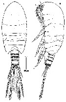 issued from : S. Chullasorn, F.D. Ferrari & H.-U. Dahms in Helgol. Mar. Res., 2010, 64; [p.44, Fig.7]. Male: A-B, habitus (dorsal and left lateral, respectively).
|
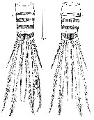 issued from : S. Chullasorn, F.D. Ferrari & H.-U. Dahms in Helgol. Mar. Res., 2010, 64; [p.45, Fig.8]. Male: A-B, urosome (dorsal and ventral, respectively).
|
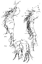 issued from : S. Chullasorn, F.D. Ferrari & H.-U. Dahms in Helgol. Mar. Res., 2010, 64; [p.46, Fig.9]. Male: A, rostrum and left A1 (anterior view); B, right A1 (anterior view); C, A2. Nota: left A1 similar to female; right A1 also 17-segmented, proximal seta on articulating segments 12-14 modified as a broad-based, spine-like seta closely appressed to segment; articulating segment 15 attenuate distally, tip reaching distal end of segment 16. A2, Mx1, Mx2 and Mxp similar to female.
|
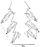 issued from : S. Chullasorn, F.D. Ferrari & H.-U. Dahms in Helgol. Mar. Res., 2010, 64; [p.47, Fig.10]. Male: P2. Lateral margin of left and right middle and distal exopod showing slight difference in length of proximal (middle) spine-like seta (arrowhead) on distal complex; imaginary broken lines for reference. Nota: P1, P3 and P4 similar to female but with denticles on basis (P1), or coxa, basis and rami (P3-P4). P2: denticles on coxa, basis and rami. Tip of proximolateral seta on left distal exopodal complex projecting beyond imaginary line between tip of lateral seta on distal exopodal complex; tip of proximolateral seta on right distal exopodal complex not reaching that imaginary line.
|
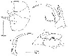 issued from : S. Chullasorn, F.D. Ferrari & H.-U. Dahms in Helgol. Mar. Res., 2010, 64; [p.48, Fig.11]. Male: A, protopod and endopods of P5 (anterior); B, right exopod of P5 (anterior); C, right bexopod of P5, medial seta of proximal segment and distal segment (posterior); D, left exopod (anterior). P5: Intercoxal plate with ventral, beak-like lobe; Right coxa fused to right basis; later with lateral seta. Right exopod 2-segmented, proximal segment with lateral seta with epicuticular fringe on both sides; broad blade-like medial seta with incomplete fringe on one side; distal segment thick, curved, spine-like attenuation with medial and lateral seta proximally. Right endopod 1-segmented, quadrate, without setae, with proximal and distal indentation. Left coxa fused to left basis; latter with lateraln seta; basis with broad attenuation narrowing and folded on itself forming grooved, pointed tip. Left exopod apparently 2-segmented, proximal segment with lateral seta with epicuticular fringe on one side, and medial attenuation with denticles; distal segment with broad, quadrate, proximolateral lobe with medial denticles, followed by small middle lobe with lateral denticles, and broad, distal lobe flexed 90°; with 2 pointed lobes apically; 1 small seta proximal to quadrate lobe and 1 small seta proximal to flexed lobe. Left endopod 1-segmented, rounded, without setae, with distal, tongue-like lobe.
| | | | | NZ: | 1 | | |
|
Distribution map of Pseudocyclops schminkei by geographical zones
|
| | | | Loc: | | | NW Pacif. (Zamami Island, Okinawa) | | | | N: | 1 | | | | Lg.: | | | (1105) F: 0,55-0,63; M: 0,56-0,59; {F: 0,55-0,63; M: 0,56-0,59} | | | | Rem.: | in aquarium used to culture pearl oysters. | | | Last update : 02/04/2019 | |
|
|
 Any use of this site for a publication will be mentioned with the following reference : Any use of this site for a publication will be mentioned with the following reference :
Razouls C., Desreumaux N., Kouwenberg J. and de Bovée F., 2005-2026. - Biodiversity of Marine Planktonic Copepods (morphology, geographical distribution and biological data). Sorbonne University, CNRS. Available at http://copepodes.obs-banyuls.fr/en [Accessed January 22, 2026] © copyright 2005-2026 Sorbonne University, CNRS
|
|
 |
 |


