|
|
 |
|
Cyclopoida ( Order ) |
|
|
|
Oncaeidae ( Family ) |
|
|
|
Triconia ( Genus ) |
|
|
| |
Triconia constricta Wi, Böttger-Schnack & Soh, 2012 (F,M) | |
| | | | | | | Ref.: | | | Wi & al., 2012 (p.844, figs.F,M, Rem.); Soh & al., 2013 (p.121, figs.F, M) | 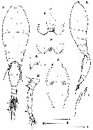 Issued from : J.H. Wi, R. Böttger-Schnack & H.Y. Soh in J. Crustacean Biol., 2012, 32 (5). [p.846, Fig.2]. Female (from 32°00'N, 126°8'E): A-B, habitus (dorsal and lateral, respectively; caudal setae numbered using Roman numerals); C, genital double-somite (dorsal); D, A1; E, labrum (anterior); F, labrum (posterior; arrow indicates small denticles on outer lateral margin); G, pleural area of 4th pedigerous omite (left side); H, P5 (left side). Nota: Prosome 1.5 times length as long as urosome including caudal rami. Proportional lengths of urosomites and caudal rami 12.5 : 54.3 : 8.7 : 6.5 : 9.8 : 5.2 = 100. Dorsal surface of genital double-somite with single pore anterior quarter and paired pores at posterior third. Paired genital apertures about 2/5 distance from anterior margin of dorsal surface, armed with spine and small spinous process near base of spine. Anal somite about 1.2 times as long as wide. Caudal ramus 1.6 times as long as wide; bearing 6 setae (setae II, III spiniform, unipinnate along medial margin, other setae IV-VII setiform and plumose. A1 6-segmented, relative lengths of segments (measured along posterior non-setiferous margin) 9.8 : 17.9 : 43.9 : 13.8 : 5.7 : 8.9. Labrum bilobed. Outer margin of each lobe with row of short spinules (indicated by arrow in fig.2F), inner part of distal margin of lobe with 6 strong denticles; medial concavity covered anteriorly by wrinkled hyaline lamella (fig.2E), anterior surface without integumental pockets and spinular patches. Posterior wall of medial concavity with 2 long and distinctly sclerotized dentiform processes and 2 short flat elements (fig.2F), posterior surface of each lobe with 2 secretory pores. P5 with small free segment representing exopod, and very long outer basal ( = protopodal) seta reaching beyond paired secretory pores on genital double-somite (arrowed in fig.2A). Exopod with long outer seta, reaching almost as far as genital apertures, and short inner seta. P6 represented by external operculum closing off each genital aperture, armed with spine and small spinous process.
|
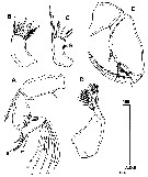 Issued from : J.H. Wi, R. Böttger-Schnack & H.Y. Soh in J. Crustacean Biol., 2012, 32 (5). [p.847, Fig.3]. Female: A, A2 (individual elements on lateral margin of 2nd endopodal segment numbered using Roman numerals, distal elements designated using capital lettes); B, Md (individual elements designated using small form letters); C, Mx1; D, Mx2; E, Mxp. Nota : A2 3-segmented, relative lengths of segments 41 : 32 : 27 = 100. Coxobasis armed with bipinnate seta at inner distal corner, protruded area on outer margin ornamented with small denticles (indicated by arrow in fig.3A). Proximal endopodal segment ornamented with row of denticles along inner margin and spinular row of convex outer margin ; armature arranged in mid-segment group of 4 elements (seta III spiniform and pectinate, setae I, II and IV naked, seta I shortest), and distal group of 7 elements (setae A-D unipinnate, setae E, F, G unornamented, seta G about 2/3 the length of seta F). Md represented by flattened gnathobase with 5 elements (ventral seta a stout, with row of long setules along dorsal margin, these gradually shortening distally ; ventral blade b broad and tapering distally, ornamented with row of short spinules on posterior surface ; dorsal blade c almost as long as ventral blade b, with several dentiform processes along entire dorsal and distal margins ; setae d short and bipinnate ; seta e longer than seta d and setose). Mx1 weakly bilobed. Inner lobe ( = praecoxal arthrite) with 3 setae unequal in length (innermost seta shortest, sparsely ornamented with spinules, located at some distance from others ; outermost seta strong, and longer than other 2 setae, fringed with few spinules. Outer lobe with 4 setae (outermost seta setiform and pectinate, longer than the other 3 setae ; seta next to innermost seta unipinnate ; innermost seta naked and shortest). Mx2 2-segmented, comprising syncoxa and allobasis. Allobasis produced distally into slightly curved claw with 2 rows of very strong spinules along medial margin ; outer slender seta extending to just below the tip of allobasal claw ; proximal medial margin with naked seta and curved spine with 2 rows of spinules along medial margin and single row of slender spinules along lateral margin. Mxp 4-segmented, syncoxa unarmed. Basis robust, with 2 spiniform, bipinnate elements on palmar margin, distal one about 1.5 times as long as proximal one ; surface of basis ornamented with fringe of fine spinules between distal seta and articulation with endopod and row of short spinules between proximal and distal setae. Proximal endopdal segment unarmed. Distal endopodal segment drawn out into long curved claw, with strong spinules along entire concave margin, with small naked seta on outer proximal margin and unipectinate spine basally fused to inner proximal corner of claw.
|
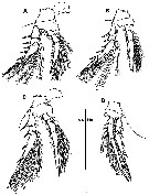 Issued from : J.H. Wi, R. Böttger-Schnack & H.Y. Soh in J. Crustacean Biol., 2012, 32 (5). [p.849, Fig.4]. Female: A, P1 (anterior view); B, P2 (anterior view, intercoxal sclerite not shown); C, P3 (anterior view); D, P4 (anterior view, intercoxal sclerite not shown).
|
 Issued from : J.H. Wi, R. Böttger-Schnack & H.Y. Soh in J. Crustacean Biol., 2012, 32 (5). [p.848]. Female: Armature formula of P1 to P4 ( Roman numerals = spines; Arabic numerals = setae).
|
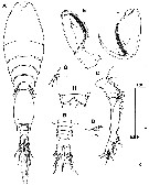 Issued from : J.H. Wi, R. Böttger-Schnack & H.Y. Soh in J. Crustacean Biol., 2012, 32 (5). [p.850, Fig.5]. Male: A, habitus (dorsal); B, anal somite and caudal rami (dorsal); C, A1; D, A2 (setae on mid-segment of distal endopodal segment); E, Mxp (anterior); F, Mxp (posterior; syncoxa omitted, arrow indicating spine basally fused to inner proximal corner of claw); G, P5 (left side, dorsal); H, P6 (arrows indicate small denticles on posterolateral margins). Nota : Proportional lengths of urosomites including caudal rami 8.6 : 57.6, 5.7 : 4.6 : 4.6 : 10.9 : 8.0 = 100. A1 4-segmented. Distal segment corresponding to fused 4th to 6th segments of female. P5 with exopod fused to 1st urosomal somite, with 2 setae different in length (outer seta slightly longer than inner one). Protopodal seta slightly longer than outer exopodal setae. P6 represented by posterolateral flap closing off genital aperture on either side; distal margin of genital flap ornamented with row of denticles (arrowed in fig.5H); posterlateral corners sharply protruding.
|
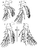 Issued from : J.H. Wi, R. Böttger-Schnack & H.Y. Soh in J. Crustacean Biol., 2012, 32 (5). [p.851, Fig.6]. Male: A, P1 (anterior view); B, P2 (anterior view, intercoxal sclerite not shown); C, P3 (anterior view); D, P4 (anterior view, intercoxal sclerite not shown).
|
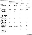 Issued from : J.H. Wi, R. Böttger-Schnack & H.Y. Soh in J. Crustacean Biol., 2012, 32 (5). [p.852, Table 1]. Comparison of morphological features of females of three species of the dentipessubgroup from the Red Sea and Triconia constricta n. sp. from the East China Sea. * Calculated after Böttger-Schnack (1999: table 2, figs. 9, 11, 27, 29, 31, 33). CR = caudal rami.
|
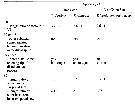 Issued from : J.H. Wi, R. Böttger-Schnack & H.Y. Soh in J. Crustacean Biol., 2012, 32 (5). [p.852, Table 1]. Comparison of morphological features of males of two species of the dentipessubgroup from the Red Sea and Triconia constricta n. sp. from the East China Sea. * Calculated after Böttger-Schnack (1999: table 2, figs. 12, 30). CR = caudal rami.
|
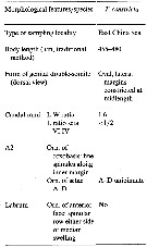 Issued from : K. Cho, W.-S. Kim, R. Böttger-Schnack & W. Lee in J. Nat. Hist., 2013. [p.16, Table 2]. Morphological characters of Triconia constricta of the Triconia dentipes-subgroup from the north-eastern equatorial Pacific. Nota: Orn: ornamentation; L: length; W: width; Compare with related species and form variants of the Triconia dentipes-subgroup from the north-eastern equatorial Pacific and from other regions: T. dentipes, T. pacifica, T. elongata, T. giesbrechti (Pacific form), T. giesbrechti (Red Sea form).
|
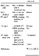 Issued from : K. Cho, W.-S. Kim, R. Böttger-Schnack & W. Lee in J. Nat. Hist., 2013. [p.17, Table 2 (Continued)]. Morphological characters of Triconia constricta of the Triconia dentipes-subgroup from the north-eastern equatorial Pacific. Nota: Orn: ornamentation; L: length; W: width; OSDS: outer subdistal spine; ODS: outer distal spine; CP: distal conical process Compare with related species and form variants of the Triconia dentipes-subgroup from the north-eastern equatorial Pacific and from other regions: T. dentipes, T. pacifica, T. giesbrechti (Pacific form), T. giesbrechti (Red Sea form), T. elongata.
| | | | | NZ: | 1 | | |
|
Distribution map of Triconia constricta by geographical zones
|
| | | | | | | Loc: | | | East China Sea (S Jeju Island)
Locality type: 32°00'N, 126°8'E. | | | | N: | 1 | | | | Lg.: | | | (1144)* F: 0,455-0,479; M: 0,390-0,409; (1174) F: 0,46; M: 0,40; {F: 0,455-0,497; M: 0,390-0,409}
*Body length (including caudal ramus) measured in lateral view. | | | | Rem.: | For Wi & al. (2012, p.848) this species is closely related to T. dentipes, T. elongata, and T. giesbrechti, together forming the dentipes-subgroup. Among the 3 described species of the dentipes-subgroup, T. constricta is most similar to T. giesbrechti. | | | Last update : 06/04/2015 | |
|
|
 Any use of this site for a publication will be mentioned with the following reference : Any use of this site for a publication will be mentioned with the following reference :
Razouls C., Desreumaux N., Kouwenberg J. and de Bovée F., 2005-2025. - Biodiversity of Marine Planktonic Copepods (morphology, geographical distribution and biological data). Sorbonne University, CNRS. Available at http://copepodes.obs-banyuls.fr/en [Accessed December 31, 2025] © copyright 2005-2025 Sorbonne University, CNRS
|
|
 |
 |


