|
|
 |
|
Monstrilloida ( Order ) |
|
|
|
Monstrillidae ( Family ) |
|
|
|
Maemonstrilla ( Genus ) |
|
|
| |
Maemonstrilla protuberans Suarez-Morales & McKinnon, 2014 (F) | |
| | | | | | | Ref.: | | | Suarez-Morales & McKinnon, 2014 (p.328, Descr.F, figs.F, Rem.) | 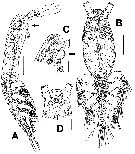 Issued from : E. Suarez-Morales & A.D. McKinnon in Zootaxa, 2014, 3779 (3). [p.329, Fig.18]. Female (from 19°7.34'S, 146°53.024'E): A, habitus showing preoral protuberance (arrow), lateral view; B, habitus (ventral view); C, cephalic area showing cuticular reticulation, preoral ventral protuberance (arrow), and oral papilla, lateral view; D, same (ventral view). Scale bars: A, B = 100 µm; C, D = 50 µm. Nota: Cephalothorax representing up to 58 % of total body length (measured from the anterior end of the cephalothorax to the posterior end of the anal somite), covered with pattern of minute spinules mixed with light reticulation. Reticulation obseved only on anterior dorsal and ventral surfaces of cephalothorax and on some antennular segments. A1 rep^resenting 35 % of total body length (measured from the anterior end of the cephalothorax to the posterior end of the anal somite) and 61 % of cephalothorax length. Oral papilla conical, ventrally projecting , located anteriorly, about 20 % of way back along ventral surface of cephalothorax. pair of relatively large ocelli present, pigment cups weakly developed, separated by about 1/2 eys diameter, unpigmented; ventral cup slightly smaller than lateral cups. Preoral surface with ventral rounded protuberance (arrowed in fig.18C) and 2 pairs of nipple-like cuticular processes with adjacent pattern of striae (fig.18C, D). Ovigerous spine relatively short, reaching to between P2 and P3, partially concealed by relatively small egg mass (fig.18 B).
|
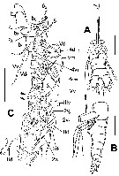 Issued from : E. Suarez-Morales & A.D. McKinnon in Zootaxa, 2014, 3779 (3). [p.330, Fig.19]. Female: A, urosome showing P5, ovigerous spines, genital pore (arrow), and caudal seta VII (arrows), dorsal view; B, same (lateral); C, left A1 showing high reticulation ridges on outer margin (arrows), dorsal view. Scale bars: A-C = 50 µm. Nota: A1 4-segmented, with weak divisions between segments 2-3 and 3-4 only marked by constrictions. High reticular ridges on outer margin of segments 3 and 4 (arrowed in fig.19C). P5 paired, rod-like, with 2 setae, 1 distal, 1 subdistal. Urosome consisting of 4 somites: 5th pedigerous somite, compound genital somite with incomplete transverse suture, and 2 free postgenital somites (i.e. preanal and anal somites). Ventral surface of genital double-somite bearing ovigerous spines arising from conical projection of ventrally protuberant anterior half. Posterior half of genital compound somite straight, without ventral process. Copulatory opening located on ventral surface at posterior base of ovigerous spine cone (arrowed in fig.19A). Spines cylindrical, smooth, straight, tapering and diverging distally, not swollen or bulbous distally. Caudal rami subquadrate, weakly divergent, approximately 1.3 times longer than wide, each ramus bearing 6 setae. Inner dorsal seta (seta VII of Huys & Boxshall, 1991, p.30) thinnest (arrowed in fig.19A).
|
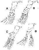 Issued from : E. Suarez-Morales & A.D. McKinnon in Zootaxa, 2014, 3779 (3). [p.331, Fig.20]. Female: A-D, P1 to P4, respectively (with intercoxal sclerites complete or cut short). Scale bars: A-D = 100 µm. Nota: Armature formula of swimming legs P1 to P4 as in M. ohtsukai.
| | | | | NZ: | 1 | | |
|
Distribution map of Maemonstrilla protuberans by geographical zones
|
| | | | Loc: | | | E Australia (Davies Reef, Queensland)
Type locality: 19°7.34'S, 146°53.024'E | | | | N: | 1 | | | | Lg.: | | | (1149)* F: 0,57; {F: 0,57}
* Body length measured from the anterior end of cephalothorax to the posterior end of the anal somite. | | | | Rem.: | After Suarez-Morales & McKinnon (2014, p.332) this species is assignable to the M. hyottoko species group as difined by Grygier & Ohtsuka (2008) mainly by its possession of a lightly reticulate cephalothorax and A1 mixed with a dense spinulose pattern, the absence of an inner seta on the 1st exopodal segment pof P1-P4, and unbranched, rod-like P5 with 2 setae. | | | Last update : 17/01/2015 | |
|
|
 Any use of this site for a publication will be mentioned with the following reference : Any use of this site for a publication will be mentioned with the following reference :
Razouls C., Desreumaux N., Kouwenberg J. and de Bovée F., 2005-2026. - Biodiversity of Marine Planktonic Copepods (morphology, geographical distribution and biological data). Sorbonne University, CNRS. Available at http://copepodes.obs-banyuls.fr/en [Accessed May 07, 2026] © copyright 2005-2026 Sorbonne University, CNRS
|
|
|
|


;)
;)
;)
