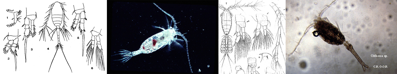
Species Card of Copepod
Calanoida ( Order )
    Pseudocyclopoidea ( Superfamily )
        Pseudocyclopidae ( Family )
            Pinkertonius ( Genus )
Pinkertonius ambiguus  Bradford-Grieve, Boxshall & Blanco-Bercial, 2014   (F,M)
Ref.:
Bradford-Grieve & al., 2014 (p.517, Descr.F,M, figs.F;M, genetic catalogued in GenBank); Laakmann & al., 2019 (p.330, fig. 2, 3, phylogenetic relationships)
Species Pinkertonius ambiguus - Plate 1 of morphological figuresIssued from : J.M. Bradford-Grieve, G.A. Boxshall & L. Blanco-Bercial in Zool. J. Linn. Soc., 2014, 171. [p.511, Fig.2].
Female (44°41.95'S, 173°41.94'E): A-B, habitus (dorsal and lateral, respectively); C, A1; D, rostrum; E, genital double-somite (ventral); F, caudal rami (dorsal); G, same (ventral); H, P5 (anterior); I, detail of joint between exopod segments 2 and 3 of P5, showing (large, unlabelled arrow-heads) location of two pivot points. Selected characters and their states are indicated (e.g for example arrowhead 5.2).
Scale bars: A-C, 1.0 mm (A1 at same scale as A and B); D-I, 0.1 mm.

Nota : Cephalosome and pedigerous segment 1 separate, 4th and 5th separate.
Pedigerous somite 5 produced into pointed lappets extending more than ½ along genital double-somite.
Rostrum in form of ventrallydirected rounded plate with pair of filaments.
Urosome 4-free somites, fist three bordered posteriorly by unevenly serrated hyaline fringe, largely intact ventrally.
Genital double-somite with slight anterior swelling in dorsal view ; in lateral view swollen ventroanteriorly ; in ventral view genital field asymmetrical, gonopores slightly unequally developed, being larger on left, genital operculum kewed to left with hinge aligned at about 45° to anterior-posterior axis of somite, so that right gonopore ant completely covered, right side of genital field bordered by ridge aligned anterior-posteriorly, no such flange on left, copulatory pore and seminal receptacle not obvious, although sac on left apparently linked to left gonopore.
Caudal rami slightly asymmetrical, longer on right, with 7 setae each. Seta I vestigial, seta II spiniform, seta V longest, seta IV next longest followed by setae VI and III, seta VII small and spiniform and inserted on dorsoinner distal corner ; left caudal ramus inner border lined with fine setules ; on right ramus row of long setules arranged obliquely on anterior part of ventral surface.
A1 27-segmented, extending to posterior border of pedigerous somite 5.
P5 articulation of exopodal segment 3 with segment 2, oblique (reminiscent of sepcies of Ridgewayia) ; proximal end of segment 3 narrowing, distance between two pivot points (fig.2H) ensures region of articulation is as wide as exopodal segment 3 at level of proximal inner seta of exopodal segment 3. Outer distal extension of exopodal segment 2 not reaching origin of proximal outer spine of exopodal segment 3.


Species Pinkertonius ambiguus - Plate 2 of morphological figuresIssued from : J.M. Bradford-Grieve, G.A. Boxshall & L. Blanco-Bercial in Zool. J. Linn. Soc., 2014, 171. [p.512, Fig.3].
Female: A, A1 ancestral segments I-XII; B, A1 ancestral segments XIII-XIX; C, A1 ancestral segments XX-XXVIII; D, Mx2; E, Mx2 endopod.
Scale bars: A-D, 0.1 mm; E, 0.02 mm.
Selected characters and their states are indicated (e.g for example arrowhead 20.2).

Nota : A1 with 1 aesthetasc present on each of segments I, III, VII, XI, XIV, XVI, XVIII, XXI, XXV, and XXVIII ; small cuticular thickenings found on segmentsII-X, XII, XIII, XV, and XVII ; 1 seta on eight proximal segments, wider than other setae, and attenuated distally into curved narrow tip.
Mx2 : Praecoxa, coxa and basis clearly separated, endites 1-4 with 7 (1 very short), 3, 3 and 3 setae, respectively ; basal endite with 3 setae, one of them stout and spiniform ; inner setae on endites 2-5 lined with long spinules ; endopod segment 1 endite with 3 setae, segments 2-4 with 2, 2, 3 setae, respectively.


Species Pinkertonius ambiguus - Plate 3 of morphological figuresIssued from : J.M. Bradford-Grieve, G.A. Boxshall & L. Blanco-Bercial in Zool. J. Linn. Soc., 2014, 171. [p.514, Fig.4].
Female: A, A2; B, Md; C, Mx1; D, Mxp.
Scale bars: 0.1 mm.
Selected characters and their states are indicated (e.g for example arrowhead 66:1).

Nota : A2 Coxa and basis separate ; endopod 2-segmented with traces of fusion between segments 2 and 3, and between segments 3 and 4 ; segment 1 with 2 inner setae ; segment 2 with 9 plus 7 terminal setae and outer transverse rox of spinules marking boundary between putative endopodal segments 3 and 4. Exopod shorter than endopod, 8-segmented, ancestral segments VIII and IX fused, segments I-VII each with 1 well developed seta, compound distal segment VIII-IX with 3 terminal setae and 1 inner subterminal seta, outermost seta on terminal endopodal segment lined proximally with small spinules.
Md : Gnathobase with 7 marginal teeth, ventralmost largest, 5 dorsal teeth bicuspid, small spinulated seta inserted dorsally.Basis with 4 apparently naked setae ; endopod 2-segmented segment 1 with inner lobe and 4 setae, segment 2 with 10 terminal setae, distoinner border with short row of spinules at about midlength. Exopod 5-segmented with setal formula 1, 1, 1, 1, 2.
Mx1 : Praecoxal arthrite with 14 spines and setae, including 4 on posterior surface and 1 on dorsal surface ; coxal endite with 4 setae ; basal endites 1 and 2 with 4 and 5 setae, respectively. Endopod segments 1 and 2 fused, segments 2 and 3 separate, with 4, 3, and 7 setae, respectively. Exopod with 11 setae, of which 3 terminal setae short and bordered by fine setules along inner border. Basal exite without seta. Coxal epoipodite with 9 setae, of which 3 proximal setae short.
Mxp : 1st syncoxal endite with 1 seta, endites 2 and 3 with 2 and 4 spinulose setae, respectively, crescent-shaped row of fine spinules at base of endite 3 on inner surface ; endite 4 with 3 setae and small peg-like structure, and a few small spinules. Basis with 2 setulose setae and proximal border lined by spinules. Endopod well-developed, longer than basis, endopodal segment 1 apparently separate from basis, endopodal segments 1-6 with 2, 4, 4, 3, 3 plus 1, and 4 spinulose setae, respectively, outer seta of segment 6 wider and longer than adjacent seta, and terminally inserted.


Species Pinkertonius ambiguus - Plate 4 of morphological figuresIssued from : J.M. Bradford-Grieve, G.A. Boxshall & L. Blanco-Bercial in Zool. J. Linn. Soc., 2014, 171. [p.515, Fig.5].
Female: A, P1 (posterior); B, P2 (posterior); C, P3 (posterior); D, P3 coxa (anterior); E, P4 (posterior); F, P4 coxa (anterior surface).
Scale bar: 0.1 mm.
Selected characters and their states are indicated (e.g for example arrowhead 98:1).


Species Pinkertonius ambiguus - Plate 5 of morphological figuresIssued from : J.M. Bradford-Grieve, G.A. Boxshall & L. Blanco-Bercial in Zool. J. Linn. Soc., 2014, 171. [p.519, Table 4].
Setal formulae for swimming legs.
Roman numerals indicate spines; Arabic numerals indicate setae. Outer border setation listed first in each segment, with group separated by ;


Species Pinkertonius ambiguus - Plate 6 of morphological figuresIssued from : J.M. Bradford-Grieve, G.A. Boxshall & L. Blanco-Bercial in Zool. J. Linn. Soc., 2014, 171. [p.516, Fig.6].
Male: A-B, habitus (dorsal and lateral, respectively; C, right A1 (at same scale as A and B); D, caudal rami (dorsal); E0, P5 (posterior); note the deformed left endopod segment 3 that has an extra inner seta - this seta is absent in another specimen.
Scale bars: A-C: 1.0 mm; D, E: 0.1 mm.
Selected characters and their states are indicated (e.g for example arrowhead 120:1).

Nota : Urosome 5 free somites, anterior 4 somites borderd posteriorly by unevenly serrated hyaline fringe.
Caudal rami symmetrical, with 7 setae each : seta I vestigial, seta II spiniform, seta V longest, seta IV next longest followede by setae VI and III, weta VII small and spiniform and inserted on dorsoinner distal corner, inner borders of caudal rami lined with fine setules.
A2, Md, Mx1, Mx2, Mxp, and P1 – P4 identical to those of female.
P5 biramous on both sides, with both rami 3-segmented, asymmetrical. Exopod slightly longer on right. Right exopodal segment 2 with triangular inner attenuation, segment 3 in form of claw with terminal spine, rounded distally, fused to segment, with 2 articulated spines (1 medioproximal, other on outer border more distally inserted), with additional outer fused spinule and pore opening. Left leg exopodal segment 2 with inner border swollen, bearing seta modified into scalpel-shaped element thickened along its outer border ; exopodal segment 3 simple,about twice as long as wide, short terminal spine fused to segment at level adjacent to lateral pore opening, 1 articulated inner spine an douter articulated spine more proximally inserted on posterior surface. Posterior surfaces of exopods and endopods ornamented with scattered patches of very small spinules.


Species Pinkertonius ambiguus - Plate 7 of morphological figuresIssued from : J.M. Bradford-Grieve, G.A. Boxshall & L. Blanco-Bercial in Zool. J. Linn. Soc., 2014, 171. [p.520, Fig.7].
Male left A1: A, ancestral segments I-XIV; B, ancestral segments XV-XIX; D, ancestral segments XX-XXV; E, ancestral segments XXVI-XXVIII.
Scale bar: 0.1 mm.

Nota : Left A1 26-segmented.


Species Pinkertonius ambiguus - Plate 8 of morphological figuresIssued from : J.M. Bradford-Grieve, G.A. Boxshall & L. Blanco-Bercial in Zool. J. Linn. Soc., 2014, 171. [p.521, Fig.8].
Male right A1: A, ancestral segments I-XIV; B, ancestral segments XV-XIX; C, ancestral segments XX-XXV; D, ancestral segments XXVI-XXVIII.
Scale bar: 0.1 mm.
Selected characters and their states are indicated (e.g for example arrowhead 39:1).

Nota : Right A1 25-segmented, extending just beyond posterior border of 4th pedigerous somite, geniculation between ancestral segments XX and XXI, segments XV-XIX enlarged

NZ: 1

Distribution map of Pinkertonius ambiguus by geographical zones
Loc:
SW Pacif. (E New Zealand)

Type locality: 44°41.95'S, 173°41.94'E.
N: 1
Lg.:
(1156) F: 1,8-2,0; M: 1,8-1,88; {F: 1,8-2; M: 1,8-1,88}
Rem.: Hyperbenthic. The specimens of the new species were sorted from samples taken by an epibenthic Benke sledge deployed on the flanks of the Chatham Rise above the sea floor.
For Bradford-Grieve & al. (2014, p.517 ) the specific name ambiguus derives from the Latin, referring to the ambiguous nature of the morphology of this species in not exactly fitting into any of the previously described families. The species retains a number of plesiomorphic features: the female genital double-somite has a genital operculum; the caudal rami have a vestigial seta I; the female A1 has all of ancestral segments I-XXVII separated; all swimming legs have both rami on each side 3-segmented; male P5 of relatively simple construction; the proximal 7 segments of the A2 exopod separate; the endopod of the Md palp 2-segmented, well-developed, and bears 10 terminal setae on segment 2; Mx1 with 9 setae on the coxal epipodite,4 setae on the posterior surface of the praecoxal arthrite, and 11 exopod setae; on Mx2, endopodal segment 1 and its endite separated from the basis; Mxp endopodal segment 1 separated from the basis, and endopodal segment 5 with an outer seta.
With inclusion of this species the essay of bibliographic overview has come to an end. The work is made freely available to all who are faced with the identification and ecological aspects of marine pelagic copepods (or those from extreme habitats), occurring throughout the oceans and seas of the planet. The cited and analysed references are present in the library of the "Observatoire Océanologique de Banyuls-sur-Mer", France. This work is meant to pay tribute to all those who have contributed to the knowledge of this group of marine organisms and to the first zoogeographical review established by R. B. Seymour Sewell (Lieutenant-Colonel I.M.S.).
Last update : 02/04/2019
    to add your remark
  

 Any use of this site for a publication will be mentioned with the following reference :

Razouls C., Desreumaux N., Kouwenberg J. and de Bovée F., 2005-2024. - Biodiversity of Marine Planktonic Copepods (morphology, geographical distribution and biological data). Sorbonne University, CNRS. Available at http://copepodes.obs-banyuls.fr/en [Accessed March 29, 2024]

© copyright 2005-2024 Sorbonne University, CNRS

Webmaster
CNRS   Observatoire Océanologique de Banyuls sur Mer - Laboratoire Arago
Sorbonne Université


Version française
English version


Search

On the WEB of CNRS


Marine Planktonic Copepods

Marine Planktonic Copepods


Imprimer Contact Accueil Plan du site Accès restreint Retour Une du Labo Imprimer Contact Plan du site Crédits Téléchargez les Plug-Ins