|
|
 |
|
Misophrioida ( Order ) |
|
|
|
Speleophriidae ( Family ) |
|
|
|
Speleophria ( Genus ) |
|
|
| |
Speleophria nullarborensis Karanovic & Eberhard, 2009 (F) | |
| | | | | | | Ref.: | | | Karanovic & Eberhard, 2009 (p.51, Drecr. F, figs.F, Rem., spp key) | 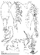 Issued from : T. Karanovic & S.M. Eberhard in Zootaxa, 2009, n° 2059. [p.54, Figs. 1-6]. Female (from 32°00'28'' S, 127°00'58'' E): 1-2, habitus (dorsal and lateral, respectively); 3, A1; 4, A2; 5, Md; 6, Mx1. Scale bars = 0.1 mm. Nota: Prosome/urosome ratio 1.7 and greatest width between 1st and 2nd free prosomites. Cephalothorax about 2.1 times as wide as genital double-somite. Posterior corners of cephalothorax and free prosomites without pronounced lateral expansions, except those of 4th free prosomite, latter somewhat expanded posterolaterally but not pointed. Urosome narrow, with 5 prosomite narrower than genital double-somite. Nauplius eye absent. Rostrum sickle-shaped from lateral aspect, much longer than 1st antennular segment, triangular with rounded tip in frontal aspect.. Prosome comprising cephalothorax and 4 free prosomites (1st pedigerous somite not incorporated into cpehalothorax), ovoid, about 2 times as long as wide (dorsal view); length ratio beginning with cephalothorax 100 : 27 : 18 : 20 : 4. Cephalothorax 1.2 times longer than its greatest width, representing 34% of total body length. Surface of cephalic shield, as well as those of free prosomites without any sensilla or pores visible. Hyaline fringe of prosomites narrow and smooth. A1 implanted on triangular pedestal, unornamented, incompletely 21-segmented, reaching posterior 3/4 of cephalothorax. Aesthetasc hypertrophied on 1st and 5th segments. A2 comprising coxa, basis, 2-segmented endopod and 6-segmented exopod (1st and 2nd exopodal segments partly fused along anterior surface). Exopod setal formula 1, 2, 1, 1, 1, 5. Md palp consisting of basis, 2-segmented endopod and indistinctly 4-segmented exopod. Coxal gnathobase cutting edge with isolated large tooth ventrally, plus row of 9 smaller, heterogeneous teeth and 2 dorsal setae. Exopod about as long as endopod, but somewhat wider, with setal formula 0, 1, 1, 4. Mx1 composed of large praecoxa, smaller coxa, large basis and 1-segmented endopod and exopod. Praecoxa with massive arthrite, armed with 2 slender setae on posterior surface and 12 elements along inner margin (9 spines and 3 setae). Coxa with well developed endite, bearing 2 spiniform and 3 slender setae; epipodite small, with 8 plumose setae. Basis with single slender, smooth outer seta; proximal endite well developed, somewhat longer than coxal endite, armed with 4 setae (3 pinnate, one of which is strong; 1 slender and smooth); distal endite indistinct, armed with 4 smooth setae. Endopod half as long as basis, armed with 5 smooth setae along inner margin and cluster of 6 setae apically. Exopod slightly larger than basis (excluding endites), twice as large as endopod, armed with 2 outer, 4 apical and 3 inner plumose setae. Exopod ornamented with long spinules along both inner and outer margins; basis ornamented only along dorsal side; other segments unornamented.
|
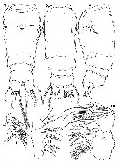 Issued from : T. Karanovic & S.M. Eberhard in Zootaxa, 2009, n° 2059. [p.56, Figs. 7-12]. Female: 7-9, Abdomen (ventral, dorsal and lateral, respectively); 10, Mx2; 11, syncoxa and basis of Mxp; 12, endopod of Mxp. Scale bar = 0.1 mm. Nota: Urosome 5-segmented, with genital and 1st abdominal somites completely fused to form double-somite. 1st urosomite ornamented with 2 large dorsal sensilla and smooth fringe dorsally and ventrally. Sclerotized joint between 1st urosomite and genital double-somite well developed, almost as pseudosomite, but weakly sclerotized dorsally. Genital double-somite about 1.5 times as long as wide (dorsal view), ornamented with 2 lateral and 2 dorsal sensilla at 1/3 and 4 posterolateral sensilla (2 on each side). Single midventral copulotary pore relatively large, almost triangular when closed, situated at 3/5 of double-somite's length; copulatory duct wide, short and rigidly sclerotized, directed dorsally and then anteriorly and attached to posterior part of seminal receptacle. Receptacle seminal small, rounded, representing only 30% of double-somite's width and 22% of its length. Paired ovipores located ventrolaterally as narrow slits at each side of copulatory pore, covered by reduced P6 and connected to receptacle via weakly sclerotized, narrow receptacle ducts. 3rd urosomite ornamented with 2 posterolateral sensilla (1 on each side). 4th urosomite (preanal somite) unornamented, about 0.3 times as long as 3rd urosomite and also much narrower than anal somite. Anal somite with smooth, broad and convex anal operculum, representing 67% of somite's width and reaching its posterior margin; ornamented with 2 large dorsal sensilla and 2 posteroventral rows of minute spinules at base of each ramus. Caudal rami subcylindrical , about as long as wide, somewhat shorter than anal somite, almost parallel; representing only about 3.5% of total body length. Distal margin ventrally straight and ornamented with small spinules, without cuticular pores visible. Only additional ornamentation consisting of several minute spinules posteriorly at base of distolateral seta. Armature consisting of 7 setae: 1 dorsal, 2 lateral and 4 apical. Dorsal seta short and smooth, 1.3 times as long as ramus, inserted close to posteromedial margin, uniarticulate at base and arising from small setophore. Proximolateral seta minute, positioned ventrolaterally at 1/4 of ramus' length. Midlateral seta strong and bipinnate, twice as long as ramus and inserted laterally at 2/5 of ramus' length. Innermost apical seta strong and bipinnate, nearly twice as long as outermost apical one (also bipinnate) and 3.6 times as long as ramus. Principal apical setae broken. Mx2 6-segmented, comprising praecoxa, coxa, basis and 3-segmented endopod. Mxp slender, 8-segmented, composed of syncoxa, basis and 6-segmented endopod. Syncoxa with proximal praecoxal endite weakly discernible, distal praecoxal and both coxal endites well developed, armature formula 1, 2, 3, 3. Endopod slender and twice as long as basis; proximalmost segment shortest, distalmost segment longest, armature formula 1, 2, 2, 2, 3, 5.
|
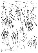 Issued from : T. Karanovic & S.M. Eberhard in Zootaxa, 2009, n° 2059. [p.59, Figs. 13-21]. Female: 13, P1; 14, 3rd exopodal segment of P1; 15, basis of P2; 16, 3rd endopodal segment of P2; 17, 3rd endopodal segment of P3; 18, 3rd exopodal segment of P3; 19, P4; 20, P5; 21, P6. Scale bars = 0.1 mm. Nota: P6 not clearly distinct, represented by trapezoidal cuticular plate, armed with inner smooth and minute spine, fused to plate, and 2 bipinnate setae; outermost seta 2.3 times as long as middle seta and 5 times as long as innermost spine.
|
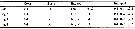 Issued from : T. Karanovic & S.M. Eberhard in Zootaxa, 2009, n° 2059. [p.57]. Female: Armature formula of swimming legs P1 to P4. Spines = Roman numerals; setae = Arabic numerals. Indicated from outer margin to inner margin.
| | | | | NZ: | 1 | | |
|
Distribution map of Speleophria nullarborensis by geographical zones
|
| | | | | | | Loc: | | | SW Australia (Nullarbor Region, Nurina cave)
Type locality: 32°00'.28''S, 127°00'58''E. | | | | N: | 1 | | | | Lg.: | | | (1168) F: 0,493; {F: 0,493} | | | | Rem.: | Anchialine cave (temperature 16.3 - 17.2°C; pH 7.38 - 6.83; salinity 30.4 - 32.1; oxygen 6.9 - 2.3 mg/L).
After Karanovic & Eberhard (2009, p.51) this species can be distinguished from its four congeners by its plesiomorphic 3-segmented endopod of P1 (2-segmented in orther species) and unusually long inermost apical seta on the caudal ramus. Another character that easily distinguishes this new species, and seems to be an autapomorphic feature (the possession of a unique derived character by a species), is its constricted preanal somite.
The discovery of this new speleophriid on the Roe Plains has important biogeographic and ecological implications. From the biogeographic perspective, it either suggests dispersal as the process determining the current distribution pattern of the aquatic fauna found on the Roe Plains or significantly extends the Tethyan track across Autralia, from the north-western coastal margin of the continent to the southern coastal margin. Vicariance model (That the components of major biota evolve together and are subject to parallel effects of geographical and climate fluctuations, and that modern biogeographical distributions result from the division of ancestral biotas by the formation of natural barriers) has been considered to be more acceptable hypothesis for explaining zoogeographic connections of subterranean faunas with disjunct distribution patterns, while dispersal model (A model explaining biogeographical distributions of whole biotas primarily in terms of dispersal of taxa across or around existing barriers) has been regarded as a better model for explaining recent distributions of marine and continental surface-water animals. | | | Last update : 05/02/2015 | |
|
|
 Any use of this site for a publication will be mentioned with the following reference : Any use of this site for a publication will be mentioned with the following reference :
Razouls C., Desreumaux N., Kouwenberg J. and de Bovée F., 2005-2026. - Biodiversity of Marine Planktonic Copepods (morphology, geographical distribution and biological data). Sorbonne University, CNRS. Available at http://copepodes.obs-banyuls.fr/en [Accessed June 27, 2026] © copyright 2005-2026 Sorbonne University, CNRS
|
|
|
|


;)
;)
;)

