Species Card of Copepod
Monstrilloida ( Order )
    Monstrillidae ( Family )
        Cymbasoma ( Genus )
Cymbasoma clauderazoulsi  Suarez-Morales, Goruppi, Olazabal & Tirelli, 2017   (F)
Ref.:
Suarez-Morales & al., 2017 (p.1811, Descr.F, figs.F, Rem.)
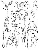
Species Cymbasoma clauderazoulsi - Plate 1 of morphological figuresIssued from : E. Suarez-Morales, A. Goruppi, A. de Olazabal & V. Tirelli in J. Nat. Hist., 2017, 51 (31-32) [p.1812, Fig.6].
Female (from Trieste): a-b, habitus (dorsal and lateral, respectively); c, cephalic region showing frontal ornamentation (dorsal); d, cephalic region, lateral view, anteriormost ventral protuberance arrowed; e, right A1 with armature (dorsal); f, urosome, dorsal view, showing posterolateral rounded processes on genital double-somite; g, urosome and P5 (setae and ovigerous spines cut short) (lateral); h, urosome and P5 (ventral); i, terminal part of the ovigerous spines (ventral); j, oral papilla and adjacent cuticular ornamentation (lateral); k, 3rd exopodal segment of P3.
Scale bars: 200 µm (a-b); 50 µm (c, d, j, k); 100 µm (e-h).

Nota : - Cephalothorax representing 64 % of total body length (caudal rami excluded)
- Midventral oral papilla located at 25 % of cephalothorax length.
- Pair of relatively small ocelli present, pigment cups moderately developed, medially separated by one eye diameter, weakly pigmented; ventral cup larger than lateral cups. Wide, dorsal symmetrical rounded protuberances surrounding ocelli (arrowed in Fig.6c) and adjacent lateral expansions in cephalic section
- Cephalic area with weakly produced forehead, ornamented.
- A pair of sensilla.
- Urosome of 3 pedigerous somites, together representing 10 % of total body length . Relative lengths of urosomites 36.1 : 41.3 : 22.6 = 100.
- Genital double-somite with smooth dorsal and ventral surfaces, with rounded posterior process on lateral margin, visible in dorsal view (arrows in Fig. 6f, g).
- Ovigerous spines paired, relatively short, representing 21 % of total body length; spine basally separated, slender, straight at their base and along shaft, without distal expansions and tapering distally, spines subequally long. Insertion of ovigerous spines with adjacent curved row of slender, short spinules.
- Caudal ramus subquadrate, armed with 3 subequally long, sparsely setulated caudal setae.
- A1 relatively long, remarkably divergent, representing about 25.6 % of thotal length and 41 % of cephalothorax length. A1 4 segmented, segments 3-4 completely fused; intersegmental division between these segments markede by elongated, relatively narrow section. Distal segment longest, representing 49 % of A1 length.
- Incorporated 1st pedigerous somite and succeding 3 pedigerous somites each bearing a pair of biramous legs. Pedigerous somites 2-4, together accounting for 17 % of total body length in dorsal view.
- P5medially conjointed, bilobate, inner (endopodal) lobe elongate, thumb-like, unarmed, rounded distally, reaching about 3/4 the length of outer lobe. Outer (exopodal) lobe elongated, slender, armed with 2 long setae on distal position plus short, slender innermost seta.

NZ: 1

Distribution map of Cymbasoma clauderazoulsi by geographical zones
Loc:
Adriatic Sea (Harbour of Trieste, Adriatic Sea )
N: 1
Lg.:
(1206)* F: 1,28; [F: 1,28] * Total length from the end of anterior cephalothorax to the posterior end of anal somite.
Rem.: For Suarez-Morales & al. (2017, p.1814) the species resembles the female of C. pseudobidentata, in the presence of: (1) - the same P5 structure and armature, with the outer lobe armed with 2 long subequal setae and a minute innermost seta; (2)- similar body proportions, with a cephalothorax representing 60-64 % of body length; (3) - diverging A1 representing 25 % of body length; (4) - a similar shape and proportion of the genital compound and anal somites, particularly with a pair of posterolateral processes. The differences between these two species are: (1) - the different A1 segmentation, with a distinctive elongated intersegmental section of segments 3-4 in the new species (Fig.5e), a character that is absent in C. pseudobidentatum (Fig.4c); (2) - A1 element 3 relatively short, thick in C. pseudobidentatum and long, slender in the new species; also, element 2v1 remarkably long in C. pseudobidentatum, longer than all the elements of group 2v-d, whereas this element is shorter than other elements of the group in the new species (Fig. 6e); (3) - the shape and size of the posterolateral processes on the genital double-somite; these are small, subtriangular in C. pseudobidentatum (Fig.4e) and larger, widely rounded in the new species (Fig.6f); (4) - the anal somite is ventrally produced in C. pseudobidentatum and flat in the new species (Fig.6g); (5) - the new species is smaller than C. pseudobidentatum and the cephalothorax shape is different in these species, the former with a narrower cephalic section and relatively smaller eyes (Fig.6a).
P5 with an inner lobe present and an outer lobe armed with 2 long setae and an inconspicuous minute inner seta is present in C. nicolettae, C. thompsonii and in records of C. rigidum (Sars, 1921; Wilson, 1932; Isaac, 1975) in which the innermost seta is slender and very small (i.e. less than half the length of the other setae). C. clauderazoulsi can be distinguisehd from this group of species by the presence of the posterior processes on the genital double-somite, but also by other characters. Among these species, only the mediterranean C. nicolettae (Suarez-Morales, 2002) has a long , divergent A1 and a similar P5 (with short innermost seta) and similar body shape and proportions; it differs from C. clauderazoulsi in the mammiliform shape of the P5 inner lobe, thus diverging from the simple thumb-like condition observed in in the new species. Also, the innermost seta is noticeably longer and more conspicuous in C. nicilettae (Suarez-Morales, 2002, Fig.8) than in the new species. The A1 structure is similar in both species, with an elongated intersegmental section between segmets 3-4, but in C. nicolettae elements 3 (short, robust) and 2v1 (shorter than other elements of group 2v-d) (Suarez-Morales, 2002, Figs. 5 and 6) differ from the elements observed in C. clauderazoulsi (both elements 3 and 2v1 are long and slender).
Last update : 19/06/2023
    to add your remark
  

 Any use of this site for a publication will be mentioned with the following reference :

Razouls C., Desreumaux N., Kouwenberg J. and de Bovée F., 2005-2024. - Biodiversity of Marine Planktonic Copepods (morphology, geographical distribution and biological data). Sorbonne University, CNRS. Available at http://copepodes.obs-banyuls.fr/en [Accessed November 21, 2024]

© copyright 2005-2024 Sorbonne University, CNRS

Webmaster
CNRS   Observatoire Océanologique de Banyuls sur Mer - Laboratoire Arago
Sorbonne Université


Version française
English version


Search

On the WEB of CNRS


Marine Planktonic Copepods

Marine Planktonic Copepods


Imprimer Contact Accueil Plan du site Accès restreint Retour Une du Labo Imprimer Contact Plan du site Crédits Téléchargez les Plug-Ins