|
|
 |
|
Monstrilloida ( Order ) |
|
|
|
Monstrillidae ( Family ) |
|
|
|
Monstrillopsis ( Genus ) |
|
|
| |
Monstrillopsis longilobata Lee, Kim & Chang, 2016 (M) | |
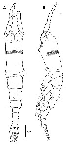
| | | | | | | Ref.: | | | Lee J. & al., 2016 (p.411, Descr. M, figs. M) |  Issued from : J. Lee, D. Kim & C.Y. Chang in Zootaxa, 2016, 4174 (1). [p.413, Fig. 1]. Male (from Mipo Bay): A-B, habitus (dorsal and lateral, respectively). Scale bar = 200 µm. Nota: Cephalothorax accounting for about 49.1 % of body length. - Forehead with one pair of sensilla near anterior margin; near posterior margin, row of 8 minute sensilla dorsally and laterally along posterior margin. - 2 groups of sensory pores situated middorsally at level of nauplius eye; anterior 3 pores aggregated near medial line, posterior 5 pores arranged in semicircle. - Nauplius eye well-developed, weakly pigmented; lateral cups set close to each other; ventral cup slightly smaller than lateral ones. - Paired sensory pores situated posterior to nipple^like processes. - Oral papilla small, locating at about 1/3 length (34 %) of cephalothorax, protruding ventrally.. - 2nd pedigerous somite ( i.e. 1st free somite) with 2 posterolateral and 1 middorsal pairs of sensilla. - 3rd pedigerous somite with 4 pairs of sensilla posteriorly and laterally. - 1 pair of pores present middorsally near anterior margin of each of pedigerous somites 2-4.
|
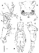
 Issued from : J. Lee, D. Kim & C.Y. Chang in Zootaxa, 2016, 4174 (1). [p.414, Fig. 2]. Male: A, anterior part of cephalothorax (ventral); B-C, urosome with genital apparatus (dorsal and lateral, respectively); D, right A1 (ventral; with setal elements labeled on the 1st to 4th segments and on the apical segment according to Huys & al.'s (2001) notation; arrow indicates a setule on inner proximal corner of the last segment according to Grygier & Ohtsuka's (1995) nomenclature, Scale bars = 100 µm. Nota: - Uraosome comprising 5th pedigerous somite, genital omite, 3 postgenital somites, (including anal somite) and caudal rami, altogether accounting for 50.9 % of total body length<. - Urosomites lacking wrinkles, except for 5th pedigerous somite and anterolateral part of genital somite. - 5th pedigerous somite small, limbless. - 4th urosomite and anal somite separated by ventral suture, not clearly visible dorsally. - Anal somite trapezoidal; lateral margins straight, with no notch or wrinkles. - Caudal rami not divergent, about 1.8 times longer than wide, swollen at basal part of inner face and with weakly concave inner margin; furnished with 4 subequal setae, comprising 1 lateral seta issuing from middle of lateral margin of ramus, 2 terminal and 1 slightly inner dorsal setae, (supposedly representing caudal setae II, IV, V and VII, respoectively of Huys & Boxshall, 1991); bases of inner terminal and lateral caudal setae heavily sclerotized, bulbous. - A1 equal to 38.7 % of total body length and 78.8 % of cephalothorax length; 5-segmented (length ratio of segments 10.3 : 25.6 : 12.6 : 26.4 : 25.4 = 100); geniculate between 4th and 5th segments, armed with one claw-like, spinous seta distally. Genital apparatus comprising relatively short shaft protruding ventrally and pair of elongate lappets. Ventral surface of shaft smooth, with no apparent protrusion or ornamentation around genital openings. - Genital lappets conjoined at base, each slightly tapering distally, nearly smooth without undulations, with round tip gently curving inward, extending slightly beyond posterior margin of anal somite, almost reaching to base of lateral caudal seta
|
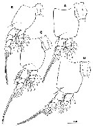
 Issued from : J. Lee, D. Kim & C.Y. Chang in Zootaxa, 2016, 4174 (1). [p.415, Fig. 3]. Male: A, P1; B, P2; C, P3; D, P4. Scale bar = 100 µm. Nota: P1-P4 with both endopod and exopod 3-segmented. Intercoxal sclerite about 1.5 times as long as wide, unornamented. Basis not divided from coxa medially; outer basal seta slender, simple, except for much longer, biserially plumose seta on P3
|
 Issued from : J. Lee, D. Kim & C.Y. Chang in Zootaxa, 2016, 4174 (1). [p.416]. Male: Armature of the swimming legs P1 to P4. Spines : Roman numerals; setae: Arabic numerals; the element or elements on the outer margin of any segment are given first.
| | | | | NZ: | 1 | | |
|
Distribution map of Monstrillopsis longilobata by geographical zones
|
| | | | | | | Loc: | | | Korea (Mipo Bay, SE coast)
Type locality: 35°31'40.71'' N, 129°26'57.68'' E. | | | | N: | 1 | | | | Lg.: | | | (1202) M: 1,69; {M: 1,69} | | | | Rem.: | For Lee J. & al. (2016, p.416) this species resembles M. chathamensis Suarez-Morales & Morales-Ramirez, 2009, and also M. sarsi Isaac, 1974, but differs by the size, transverse striation of the cephalothorax, the location of the oral papilla, heavily sclerotized and bulbous bases of caudal setae, and very long genital lappets.
After Lee J. & al. (2016, p.421), male genitalia with a relatively short median shaft with its posterolateral corners extended as elongate lappets belong subgroup ''Type II'' (Suarez-Morales & McKinnon, 2014).
M. longilobata and M. coreensis share a band of conspicuous transverse striations on the dorsal surface of the cephalothorax; although the females of these species are not known, it is expected that they might be recognizable by the unique striation band. These two species also share the characters of bulbous caudal rami and large ocelli close to each other, which are clearly different from those in the female of M. ferrarii/ Meanwhile, M. longilobata is distinguished from M. coreensis by the strongly concave and hollow distal margins of intercoxal sclerites of P1-P4 in male, which could be another diagnostic character for recogtnition of the female of M/ longilobata. | | | Last update : 19/06/2023 | |
|
|
 Any use of this site for a publication will be mentioned with the following reference : Any use of this site for a publication will be mentioned with the following reference :
Razouls C., Desreumaux N., Kouwenberg J. and de Bovée F., 2005-2026. - Biodiversity of Marine Planktonic Copepods (morphology, geographical distribution and biological data). Sorbonne University, CNRS. Available at http://copepodes.obs-banyuls.fr/en [Accessed February 13, 2026] © copyright 2005-2026 Sorbonne University, CNRS
|
|
 |
 |


