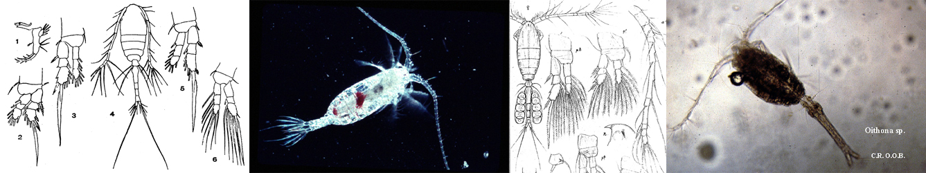
Species Card of Copepod
Calanoida ( Order )
    Clausocalanoidea ( Superfamily )
        Aetideidae ( Family )
            Pseudeuchaeta ( Genus )
Pseudeuchaeta vulgaris  Markhaseva, Mohrbeck & Renz, 2017   (F)
Ref.:
Markhaseva & al., 2017 (p.291, Descr. F, figs.F, Rem.)
Species Pseudeuchaeta vulgaris - Plate 1 of morphological figuresIssued from : E.L. Markhaseva, I. Mohrbeck & J. Renz in Mar. Biodv., 2017, 47. [p.290, Fig.1].
Female (from 11°49.8'N, 117°00.4W): a-b, habitus (dorsal and lateral, respectively); c, cephalosome (dorsal anterior); d-e, rostrum (lateral view); f, rostrum (ventral view); g, caudal rami ( (dorsal).
Scale bars: 1.0 mm (a-e); 0.5 mm (f-g).

Nota:
- Rostrum as rounded plate.
- Cephalosome and 1st pedigerous somite incpmpletely fused, 4th and 5th pedigerous somires incompletely fused.
- Posterior corners pointed directed dorsally, slightly diverging or not.
Urosome 4-segmented.
- Genital double-somite symmetrical.
- Caudal rami slightly longer than wide; 1lateral seta (II), 1 ventral seta (VII), and 4 terminal setae (III-VI).


Species Pseudeuchaeta vulgaris - Plate 2 of morphological figuresIssued from : E.L. Markhaseva, I. Mohrbeck & J. Renz in Mar. Biodv., 2017, 47. [p.291, Fig.2].
Female: a, urosome (dorsal); b-c, same (left and right lateral); d, f, h, posterior prosome and genital double-somite (lateral); e, g, i, posterior prosome and genital double-somite (dorsal); j, k, genital field in lateroventral and ventral view;
Scale bars: 1.0 mm (a-i; 0.5 mm (j-k).

Nota: Spermathecae directed dorsoanteriorly and at varying angles


Species Pseudeuchaeta vulgaris - Plate 3 of morphological figuresIssued from : E.L. Markhaseva, I. Mohrbeck & J. Renz in Mar. Biodv., 2017, 47. [p.292, Fig.3].
Female: a, A1 ancestral segments I-XI, arroxs mark small sensilla; b, A1, ancetral segments I-XVIII; c, A1, ancestral segments XIX- XXVIII; d, A1, ancestrak segments XXVII-XXVIII.
Scale bars: 1.0 mm (a-c); 0.1 mm (d).

Nota: A1 24-segmented, . longer than prosome, reaching (usually) posterior part of genital double-somite.
In some specimens small sensilla are visible on antennule proximal segments I-XI (marked by arrows).


Species Pseudeuchaeta vulgaris - Plate 4 of morphological figuresIssued from : E.L. Markhaseva, I. Mohrbeck & J. Renz in Mar. Biodv., 2017, 47. [p.293, Fig.4].
Female: a, A2; b, A2 exopod; c, Md palp; d, Md gnathobase; e, Md cutting edge; f, Mx1 (setae of proximal basal endite not figured); g-i, Mx1 praecoxal arthrite; h, Mx1proximal basal endite.
Scale bars: 0-.5 mm (a-d, f-i); 0.1 mm (e).

- A2: coxa with 1 seta, basis with 1 seta, exopod incompletely 9 segmented (2nd and 3rd proximal segments partly separate) with 1-1, 1, 1, 1, 1, 1, 1, 0, and 3 setae; 1st endopodal segment without setae, 2nd with 8+6 setae.
- Md: gnathobase cutting edge with 6 teeth, seta and needle-like spinules; basis with 1 seta; exopod 5-segmented with 1, 1, 1, 1, and 2 setae; endopod segment 1 with 1 seta, segment 2 with 5 setae (4 long and 1 additional very short and reduced un some specimens).
- Mx1: praecoxal arthrite with 9 terminal and 3 posterior setae with tiny spinules at bases of inner terminal setaz; coxal endite with 4 setae; proximal basal endite with 2 setae, distal basal endite with 4 setae (in one specimen of additional material, left limb with deviating setation of 5-6 setae); endopod with 15-16 setae; exopod with 11 setae; coxal epipodite with 7 long and 1 short setae (in one specimen of additional material, left limb with deviating setation of 9 setae), its common maxillule setation formula: 9+3, 4, 2, 4, 15-16, 11, 8.


Species Pseudeuchaeta vulgaris - Plate 5 of morphological figuresIssued from : E.L. Markhaseva, I. Mohrbeck & J. Renz in Mar. Biodv., 2017, 47. [p.294, Fig.5].
Female: a, Mx2 (praecoxal, coxal and basal endites); b, Mx2 (endite-like lobe of proximal endopodal segment and endopod; c, Mx2 (endite-like lobe of proximal endopodal segment and proximal segment of endopod; d, Mx2 (3 distal endopod segments); e, Mxp (syncoxa and proximal part of basis); f, Mxp (sensory appendage of coxa); g, Mxp (basis and endopod 'Enp'); h, Mxp (basis distal part, endopod segments 1 and 2, only 2 thick setae of endopod segment 2 figured).
Scale bars : 0.5 mm.

Nota;
- Mx2: all endites and proximal endopodal segment enditic-like lobe with 3 setae; endopod with 6 setae.
- Mxp: praecoxal endites of syncoxa with 1, 2 and 3 setae (from proximal to distal), coxal endite with 3 setae and elongated sensory attenuation; basis with 3 medial setae; endopod 6-segmented with 2, 4, 4, 3, 3+1, and 4 setae (2 setae on each of endopod 2 to 6 segments are significantly smaller and thinner); long detae of endopod segments 2-6 supplied by crescent-like structures typvcal of the genus.


Species Pseudeuchaeta vulgaris - Plate 6 of morphological figuresIssued from : E.L. Markhaseva, I. Mohrbeck & J. Renz in Mar. Biodv., 2017, 47. [p.295, Fig.6].
Female: a, P1; b, P2; c, P3; d, P4.
Scale bars: 0.5 mm.

NZ: 1

Distribution map of Pseudeuchaeta vulgaris by geographical zones
Loc:
N Pacif. (off NW Clipperton Islands

Type locality: 13°49.9' N, 116°34.5' W
N: 1
Lg.:
(1213) F: 7,00-7,60; {F: 7,00-7,60}
Rem.: Abyssal (about 4000-4400 m) samples close to the seabed.
For Markhaseva & al. (2017, p.293) the differential diagnosis of this species from its congeners is: 1- Posterior corners are pointed, directed dorsally and more or less diverging; 2- A1 reaches the middle length of posterior border of genital double-somite; 3- Md endopodal segment 2 with 5 (rarely 4) setae: 4- Mx1 coxal endite with 4 setae; proximal basal endite with 2 setae; distal basal endite with 4 setae; 5- Sensory appendage on coxal endite of Mxp syncoxa shorter than the two distal setae; 6- P1 exopodal segment 1 without lateral spine.
The species name ''vulgaris'' refers to the morphology of the species, which is typical for the genus Pseudeuchaeta.
Last update : 19/06/2023
    to add your remark
  

 Any use of this site for a publication will be mentioned with the following reference :

Razouls C., Desreumaux N., Kouwenberg J. and de Bovée F., 2005-2024. - Biodiversity of Marine Planktonic Copepods (morphology, geographical distribution and biological data). Sorbonne University, CNRS. Available at http://copepodes.obs-banyuls.fr/en [Accessed April 26, 2024]

© copyright 2005-2024 Sorbonne University, CNRS

Webmaster
CNRS   Observatoire Océanologique de Banyuls sur Mer - Laboratoire Arago
Sorbonne Université


Version française
English version


Search

On the WEB of CNRS


Marine Planktonic Copepods

Marine Planktonic Copepods


Imprimer Contact Accueil Plan du site Accès restreint Retour Une du Labo Imprimer Contact Plan du site Crédits Téléchargez les Plug-Ins