|
|
 |
|
Monstrilloida ( Order ) |
|
|
|
Monstrillidae ( Family ) |
|
|
|
Monstrilla ( Genus ) |
|
|
| |
Monstrilla sp.2 Carneiro-Schaefer, Sühnel, Dias, de Melo, Magalhaes, 2017 (Juv.F) | |
| | | | | | | Ref.: | | | Carneiro-Schaefer & al., 2017 (p.437, figs.1-6, 8-43, 14-27, Table 2, 3, Rem.) |  Issued from : A.L. Carneiro-Schaefer, S. Sühnel, C. de O. Dias, C.M. Rodrigues de Melo & A.R.M. Magalhäes in B. Inst. Pesca, Sao Paulo, 2017, 43 (3). [p.440, Figs. 3-6]. Specimens of copepodide of genus Monstrilla (female) from Perna perna culture at Palhoēa, Santa Catarina, Brazil. 3: Copepodid side view. Detail of the front feeding tubes (black asterisks). 4: Copepodid side view. Detail of the front antennules pair (black asterisk). 5: Side view ofb the metasoma and urosome copepodide. Detail of four pairs of swimming appendages (1sd, 2nd, 3nd and 4th) and double genital somite with the pair of ovigerous spines (black asterisk). 6: Dorsal view of the urosome copepoid. Details of 5th legs (black arrows) and caudal rami with 6 setae (black asterisk). Bars: 40 µm (5 and 6); 200 µm (3, 4). CF: cephalosome; MT: metasomal; UR: urosome.
|
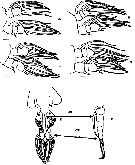
 Issued from : A.L. Carneiro-Schaefer, S. Sühnel, C. de O. Dias, C.M. Rodrigues de Melo & A.R.M. Magalhäes in B. Inst. Pesca, Sao Paulo, 2017, 43 (3). [p.441, Fig.7]. Schematic representation of swimmingt appendages pairs and urosome of copepodid Monstrilla sp.: A: P1; B: P2; C: P3; D: P4. E: ventral view of urosome indicating the 5th pair of legs (black asterisk), the pair of ovigerous spine (OS) and the caudal rami (CR).
F: side view of urosome.
Bars: 40 µm (A, B, C and D); 100 µm (E and F)
|
 Issued from : A.L. Carneiro-Schaefer, S. Sühnel, C. de O. Dias, C.M. Rodrigues de Melo & A.R.M. Magalhäes in B. Inst. Pesca, Sao Paulo, 2017, 43 (3). [p.442, Fig.16]. Longitudinal section of copepodid in the connective tissue (CT) of the mantle edge. Scale bar = 40 µm.
| | | | | NZ: | 1 | | |
|
Distribution map of Monstrilla sp.2 by geographical zones
|
| | | | Loc: | | | SW Atlant. (Santa Catarina coast, S. Brazil) | | | | N: | 1 | | | | Rem.: | For Carneiro-Schaffer & al. 2017, (p.443): In mussels culture Perna perna (from nodules located in the mussels mantle border); Mussels from commercial farms. The presence of this copepodid was recorded a prevalence of 43.33% in the culture at Palhoēa, without causing hemocyte infiltration in their hosts; this fact can be attributed to the mussels have no response to the parasite infestation. The identification of copepodids females was possible by the presence of the double-genutal somite, the ovigerous spines and 6 setae in the caudal rami. The specimens analyzed show similarities (number setae in 5th pair of leg and existing somites after genital segment with those found by Suarez-Morales & al. (2010) in Penha (SC. | | | Last update : 21/09/2020 | |
|
|
 Any use of this site for a publication will be mentioned with the following reference : Any use of this site for a publication will be mentioned with the following reference :
Razouls C., Desreumaux N., Kouwenberg J. and de Bovée F., 2005-2025. - Biodiversity of Marine Planktonic Copepods (morphology, geographical distribution and biological data). Sorbonne University, CNRS. Available at http://copepodes.obs-banyuls.fr/en [Accessed December 26, 2025] © copyright 2005-2025 Sorbonne University, CNRS
|
|
 |
 |


