|
|
 |
|
Calanoida ( Order ) |
|
|
|
Arietelloidea ( Superfamily ) |
|
|
|
Arietellidae ( Family ) |
|
|
|
Metacalanus ( Genus ) |
|
|
| |
Metacalanus sp.2 Ohtsuka, Boxshall & Roe, 1994 (F) | |
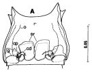
| | | | | | | Ref.: | | | Ohtsuka & al., 1994 (p.143, Descr.F, figs.F) |  issued from : S. Ohtsuka, G.A. Boxshall & H.S.J. Roe in Bull. nat. Hist. Lond. (Zool.), 1994, 60 (2). [p.139, Fig.21, A]. Female (E China Sea): A, genital double-somite (ventral). cd = copulatory duct; cp = copulatory pore; g = gonopore; rd = receptacle duct; o, oviduct; s, spermatothore remnant; sr = seminal receptacle. Scale bar in mm. Nota: Genital double-somite wider than long, symmetrical, with paired gonopores and copulatory pores located ventrolaterally near posterior end of somite; each gonopore lacking outer cuticular lateral flap, anterior half opening, covered by oval flap; copulatory pore small, round (1.4 µm in diameter) located near anterior inner corner of gonopore (spermatophore remnant attached to opening). Internal genital system similar to thaht of M. sp1. Anal operculum triangular (as in M. sp1). Cephalosome separate from 1st pedigerous somite. Lateral lobe of last prosomal somite produced backwards reaching halfway along 2nd urosomal somite. A1 asymmetrical, left longer than right, different in fusion pattern and armature. Right A1 segments X to XI, and XIV and XV only partly fused near posterior margin. Left A1 segments X and XI partly fused near posterior margin; suture between segments XI and XII visible on both surfaces, XII and XIII only on one surface, XIII and XIV completely fused. A2 with Metacalanus sp.1.
|
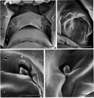
 issued from : S. Ohtsuka, G.A. Boxshall & H.S.J. Roe in Bull. nat. Hist. Lond. (Zool.), 1994, 60 (2). [p.142, Fig.24]. Female (SEM micrographs of genital double-somite): A, copulatory pores indicated by arrows); B, left gonopore and copulatory pore (indicated by an arrow); C, right copulatory pore; D, left copulatory pore. Scale bars: 20 µm (A); 10 µm (B); 2 µm (C-D).
|
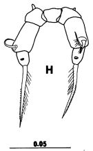
 issued from : S. Ohtsuka, G.A. Boxshall & H.S.J. Roe in Bull. nat. Hist. Lond. (Zool.), 1994, 60 (2). [p.144, Fig.26 H]. Female: H, P5 (posterior). Scale bar in mm. Nota: P5 with coxae separate from intercoxal sclerite; right basal seta thicker than left; endopod absent; right and left exopods each 1-segmented, bulbous, with spiniform seta terminally. P5 resembles that of M. curvicornis but it can be distinguished from the latter by the smaller body, the longer of A1 and by differences in the mouthparts. Legs P1 to P4 with same segmentation and setation as sp.1.
|
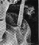
 issued from : S. Ohtsuka, G.A. Boxshall & H.S.J. Roe in Bull. nat. Hist. Lond. (Zool.), 1994, 60 (2). [p.143, Fig.25 B]. Female (SEM micrographs): B, Mandibular endopod, indicated by arrow. Scale bar = 5 µm. Nota: Mandibular palp with endopod rudimentary, 1-segmented , with 1 plumose seta; exopod with setation as in Metacalanus sp.1. Mx1: praecoxal arthrite without elements; coxal endite with short seta; coxal epipodite with 5 setae; no basal seta; endopod represented by small, unarmed knob. Mx2 and Mxp as in Metacalanus sp.1.
| | | | | NZ: | 1 | | |
|
Distribution map of Metacalanus sp.2 by geographical zones
|
| | | | | | | Loc: | | | East China Sea (off Okinawa) | | | | N: | 1 | | | | Lg.: | | | (263) F: 0,88-0,84; {F: 0,84-0,88} | | | Last update : 17/01/2015 | |
|
|
 Any use of this site for a publication will be mentioned with the following reference : Any use of this site for a publication will be mentioned with the following reference :
Razouls C., Desreumaux N., Kouwenberg J. and de Bovée F., 2005-2026. - Biodiversity of Marine Planktonic Copepods (morphology, geographical distribution and biological data). Sorbonne University, CNRS. Available at http://copepodes.obs-banyuls.fr/en [Accessed February 11, 2026] © copyright 2005-2026 Sorbonne University, CNRS
|
|
 |
 |


