|
|
 |
|
Calanoida ( Order ) |
|
|
|
Arietelloidea ( Superfamily ) |
|
|
|
Arietellidae ( Family ) |
|
|
|
Paramisophria ( Genus ) |
|
|
| |
Paramisophria itoi Ohtsuka, 1985 (F,M) | |
| | | | | | | Ref.: | | | Ohtsuka, 1985 (p.287, figs.F,M); Othman & Greenwood, 1993 (p. 86); Ohtsuka & al., 1994 (p.160, Rem.); Chihara & Murano, 1997 (p.720, Pl.: 54: F,M); Soh & al., 2013 (p.11, figs.F) | 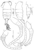 issued from : S. Ohtsuka in Publ. Seto Mar. Biol. Lab., 1985, 30 (4/6). [p.289, Fig.1]. Female (from Tanabe Bay, Honshu): A, habitus (dorsal); B, idem (lateral right side); C, urosome (lateral right side); D, right A1; E, left A1. Nota: Head and 1st thoracic segment separate, 4th and 5th fused. A1 21-segmented, right slightly longer than the left one. Urosome 4-segmented. Anal segment very small. Male: F, right A1; G, left A1. All setae and aesthetascs of A1 omitted.
|
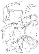 issued from : S. Ohtsuka in Publ. Seto Mar. Biol. Lab., 1985, 30 (4/6). [p.290, Fig.2]. Female: A, A2; B, endopodite of A2; C, exopodite of A2: D, Md; E, Md (mandibular cutting edge); G, Mx1. Male: F, Md (mandibular cutting edge). Nota: Basipod of A2 with a minute seta on its distal edge close to the base of endopod; endopod 2-segmented, proximal segment with a setula, distal with 3 setae and, 1 short and5 long setae apically, each segment furnished with numerous fine spinules on one side; exopod 6-segmented, first three segment almost fused, 3rd, 4th and 5th with 1, 2, 1 setae, respectively. Basipod of Md with a hairy row on inner margin and very fine spinules on outer margin; endopod wanting; exopod 5-segmented with 1, 1, 1, 1 and 2 plumose setae. Gnathobase (1st inner lobe) of Mx1 with 4 stout spines and one short process, 2nd inner lobe without seta, outer lobe with with 8 plumose setae; endopod bulb-like with 2 minute setae terminally; exopod leaf-like with 3 plumose setae on the tip.
|
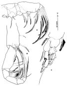 issued from : S. Ohtsuka in Publ. Seto Mar. Biol. Lab., 1985, 30 (4/6). [p.291, Fig.3]. Female: A, Mx2; B, Mxp; C, P1 (anterior). Nota: Mx2 with 6 lobes on the basal segments, 1st lobe with 1 seta, 2nd with 2 setae, 3rd and 4th each with 2 spinulose setae, 5th with a large naked spine, 6th with a serrated strong seta; endopod 3-segmented, 1st segment with 1 minute serrate and 2 serrated strong setae, 2nd and 3rd each with 2 serrated strong setae. Md 7-segmented, 3rd segment, the terminal endopodite segment with three kinds of setae: 1 naked, 1 spinulose and 2 serrated setae. with a row of ''teeth\".
|
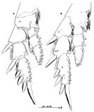 issued from : S. Ohtsuka in Publ. Seto Mar. Biol. Lab., 1985, 30 (4/6). [p.292, Fig.4]. Female: A, P2 (posterior); B, P3 (anterior).
|
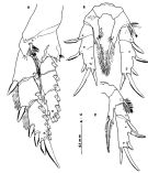 issued from : S. Ohtsuka in Publ. Seto Mar. Biol. Lab., 1985, 30 (4/6). [p.293, Fig.5]. Female: A, P4 (posterior); B, P5 (anterior); C, right P5 (anterior).
|
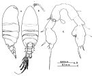 issued from : S. Ohtsuka in Publ. Seto Mar. Biol. Lab., 1985, 30 (4/6). [p.295, Fig.6]. Male: A, habitus (lateral right side); B, idem (dorsal); C, P5 (posterior). Nota: Right A1 21-segmented, first eight segments with long stout hairs on the inner margin, 4th and 5th segments partly fused together; left A1 19-segmented modified to a poorly developed grasping organ, with long stout hairs on the inner margin of first two segments. P5 asymmetrical with a common basipodite 1; left leg with the basipodite 1 disregarded, 4-segmented; basipodite 2 possessing a small bulbous rudimentary endopodite on the middle of the inner margin line at the level of a quarter the length of the terminal segment (the second fusion line is not visible in all specimens. Urosome 5-segmented.
|
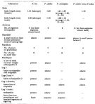 issued from : S. Ohtsuka in Publ. Seto Mar. Biol. Lab., 1985, 30 (4/6). [p.298, Table 2]. Comparison of three species within the genus Paramisophria
|
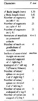 Issued from : B.-J. Lim & G.-S. Min in J. Nat. Hist., 2014, 48 (9-10). [p.532, Table 3]. Morphological characteristics. Nota: Compare within Paramisophia species of northeastern Asia: See P. sinjinensis, P. platysoma, P. japonica, P. sinica, P. koreana.
|
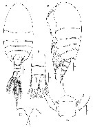 Issued from : H.Y. Soh, S.Y. Moon & J.H. Wi in Invertebrate Fauna of Korea (eds) Incheon: NIBR, 2013, 21 (28). [p.12, Fig.2]. Female (from Korean waters): A-B, habitus (dorsal and lateral, respectively); C, forehead (frontal view); D, posterior part of prosome and genital segment (dorsal); E, posterior segments of urosome (dorsal). A, B = 200 µm; C-E = 100 µm.
|
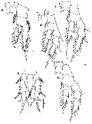 Issued from : H.Y. Soh, S.Y. Moon & J.H. Wi in Invertebrate Fauna of Korea (eds) Incheon: NIBR, 2013, 21 (28). [p.13, Fig.3]. Female: A, P1; B, P2; C, P3; D, P4; E, P5. Scale bars: 100 µm.
| | | | | NZ: | 1 | | |
|
Distribution map of Paramisophria itoi by geographical zones
|
| | | | Loc: | | | Japan (Tanabe Bay), Korea waters. | | | | N: | 2 | | | | Lg.: | | | (291) F: 1,61; M: 1,3; (1174) F: 1,83; {F: 1,61-1,83; M: 1,30} | | | | Rem.: | hyperbenthic (depth: 5 m).
For Ohtsuka (1985, p.298) , as shown in Table 2, Tanaka's materials differ from P. cluthae described by Scott (1897) in some characteristics of Md and Mx1, as ponted out by Fosshagen( 1968) | | | Last update : 03/04/2015 | |
|
|
 Any use of this site for a publication will be mentioned with the following reference : Any use of this site for a publication will be mentioned with the following reference :
Razouls C., Desreumaux N., Kouwenberg J. and de Bovée F., 2005-2025. - Biodiversity of Marine Planktonic Copepods (morphology, geographical distribution and biological data). Sorbonne University, CNRS. Available at http://copepodes.obs-banyuls.fr/en [Accessed June 04, 2025] © copyright 2005-2025 Sorbonne University, CNRS
|
|
|
|




;)
;)
;)
;)
;)
;)
;)
;)
;)
;)