|
|
 |
|
Calanoida ( Order ) |
|
|
|
Arietelloidea ( Superfamily ) |
|
|
|
Arietellidae ( Family ) |
|
|
|
Paramisophria ( Genus ) |
|
|
| |
Paramisophria japonica Ohtsuka, Fosshagen & Go, 1991 (F,M) | |
| | | | | | | Ref.: | | | Ohtsuka & al., 1991 (p.794, figs.F,M); Ohtsuka & al., 1994 (p.135, 160,161, Rem.F, figs.F); Chihara & Murano, 1997 (p.720, Pl.: 54: F,M) | 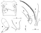 issued from : S. Ohtsuka, G.A. Boxshall & H.S.J. Roe in Bull. nat. Hist. Lond. (Zool.), 1994, 60 (2). [p.136, Fig.19]. Female (from off Okinawa): A, genital double-somite (ventral); B, exopod of A2; C, 1st and 2nd praecoxal endites of Mx2; D, 4th and 5th endopod segments of Mxp; E, 6th endopod segment of Mxp. cd = copulatory duct; cp = copulatory pore; g = gonopore; rd = receptacle duct; o, oviduct; s, spermatophore remnant; sr = seminal receptacle. Nota: Genital double-somite wider than long, with pair of gonopores anteroventrally and single copulatory pore ventromedially; seminal receptacle located lateromedially; copulatory duct thin. Mx2 with praecoxal endite with 1 seta and vestigial element, 2nd with 2 finely spinulose setae; basal spine naked. Mxp with 4th and 5th segments with relatively long innermost seta, 6th segment with setae (a and b) not reduced Scales in mm.
|
 issued from : S. Ohtsuka, G.A. Boxshall & H.S.J. Roe in Bull. nat. Hist. Lond. (Zool.), 1994, 60 (2). [p.137, Fig.20]. Female: F, P5 (anterior). Nota: P5 with coxae and intercoxal sclerite almost completely fused to basis with fine suture visible on posterior surface; 1st exopod segment separate from 2nd, 2nd and 3rd exopod segments fused. Scale bar in mm.
|
 issued from : S. Ohtsuka, A. Fosshagen & A. Go in Zool. Sci., 1991, 8 [p.794, Fig.1]. Female (from off Kume Is;, Okonawa): A, habitus (dorsal); B, idem (lateral right side); C, forehead (lateral); D-F, pediger 5 (lateral left side); G, idem (lateral right side); H, genital segment (ventral); I, anal segment and caudal rami (dorsal); J, urosome with spermatophore (lateral right side); K, genital segment with spermatophore (ventral). Holotype: A, B, D, H, I; paratypes: C, E-G, J, K. Nota: Cephalosome and 1st pediger segment separated, 4th and 5th fused. Rostrum with a pair of subterminal filaments. Dorsolateral lobe and dorsal process of pediger 5 are slightly variable in shape. Urosome 4-segmented, one-third the length of prosome. Genital segment with a pair of ventrolateral gonopores anteriorly; two small copulatory openings located closely together at middle part of ventral surface and connecting with sigmoid seminal receptacle of each side through fine tube. Anal segment small. The spermatophore on the ventral side of the genital segment covers the copulatory pores (see details in fig.5)
|
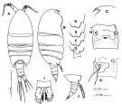
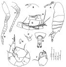
 issued from : S. Ohtsuka, A. Fosshagen & A. Go in Zool. Sci., 1991, 8 [p.795, Fig.2]. Female: A, left A1; B, right A1; C, A2; D, Md (mandibular cutting edge); E, Md (mandibular palp); F, Mx1; G, Mx2; H, Mxp. Nota: Left A1 longer than right; both A1 22-segmented. A2 with endopod 2-segmented; exopod 6-segmented (proximal two segments incompletely fused), terminal segment with 1 medial seta, and 2 setae and 1 minute setule terminally. Md: Mandibular palp with rudimentary endopod 1-segmented, bearing 2 setae of unequal lengths terminally; exopod 5-segmented. 1st inner lobe (gnathobase) of Mx1 with 5 spines and one process, 2nd inner lobe with rudimentary minute setule on tip; 1st outer lobe bearing 8 setae; inner margin of basipod 2 bearing 2 rows of hairs and 1 minute middle seta; endopod 1-segmented, bulbous, and having 3 terminal setae of unequal lengths; exopod fused with basipod segment 2, bearing 3 plumose setae terminally. Mx2 with 6 weakly developped inner lobes, 5th inner lobe furnished with 1 naked strong spine; endopod segments bearing 7 large pectinate setae
|
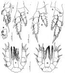 issued from : S. Ohtsuka, A. Fosshagen & A. Go in Zool. Sci., 1991, 8 [p.796, Fig.3]. Female: A, P1 (anterior); B, terminal segment of endopod of P1 (anterior); C, P2 (anterior); D, P3 (anterior); E, P4 (posterior); F, P5 (anterior); G, P5 (posterior). Holotype: A-F; paratype: G. Nota: A fusion line between the endopod and basipod 2 of P5 is not visible from both sides in the holotype , whereas it is clarly visible on the posterior surface in a paratype.
|
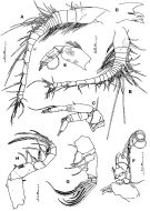
 issued from : S. Ohtsuka, A. Fosshagen & A. Go in Zool. Sci., 1991, 8 [p.797, Fig.4]. Male: A, habitus (lateral left side); B, forehead (lateral); C, pediger 5 and urosome (lateral right side); D, pediger 5 and urosome (dorsal); E, left A1 (all setae omitted); F, right A1 (all setae omitted); G, left A1 (segments 16-20); H, left A1 (segments 16-17, ventral view); I, P5 (anterior; short, thick spinule on outer process indicated by arrowhead); J, terminal segments of right P5 (posterior); K, endopod of left P5. Nota: Urosome 5-segmented, 5th small. Left A1 geniculate, 21-segmented (segments 18 and 19 incompletely fused; segment 16 with cuticular ridge along anterior margin, and bearing large spine anteroterminally). Right A1 22-segmented, shorter than left. In right P5, the outer lamellous projection on the terminal segment is triangular in one paratype and smoothly rounded in another paratype.
|
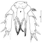
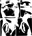 issued from : S. Ohtsuka, A. Fosshagen & A. Go in Zool. Sci., 1991, 8 [p.798, Fig.5]. Female (SEM microphotographs): A, genital segment (ventral; g = gonopore, c = copulatory pore); B-C, copulatory pores; D, right gonopore; E, left gonopore. Nota: The gonopore has an operculum with its anterior part open, and that the copulatory pores open closely in a shallow common hole located centrally on the ventral side. A paratype has a sausage-like spermatophore on the ventral side of its genital segment, which covers the copulatory pores (see fig.1, J-K)
|
 Issued from : B.-J. Lim & G.-S. Min in J. Nat. Hist., 2014, 48 (9-10). [p.532, Table 3]. Morphological characteristics. Nota: Compare within Paramisophia species of northeastern Asia: See P. itoi, P. platysoma, P. sinjinensis, P. sinica, P. koreana.
|
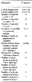
 Issued from : G. Lian & H. Qian in Acta Oceanol. Sinica, 1994, 13 (4). [p.554, Table 1]. Morphological characteristics of female from P. sinica, P. japonica and P. giselae.
| | | | | NZ: | 2 | | |
|
Distribution map of Paramisophria japonica by geographical zones
|
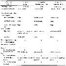
| | | | | | | Loc: | | | Japan (Tanabe Bay, Kanayama Bay), East China Sea (off Okinawa) | | | | N: | 2 | | | | Lg.: | | | (297) F: 2,08-1,85; M: 1,64-1,41; {F: 1,85-2,08; M: 1,41-1,64} | | | | Rem.: | hyperbenthic. | | | Last update : 27/01/2015 | |
|
|
 Any use of this site for a publication will be mentioned with the following reference : Any use of this site for a publication will be mentioned with the following reference :
Razouls C., Desreumaux N., Kouwenberg J. and de Bovée F., 2005-2026. - Biodiversity of Marine Planktonic Copepods (morphology, geographical distribution and biological data). Sorbonne University, CNRS. Available at http://copepodes.obs-banyuls.fr/en [Accessed June 10, 2026] © copyright 2005-2026 Sorbonne University, CNRS
|
|
|
|


;)
;)
;)
;)
;)
;)
;)
;)
;)

