|
|
 |
|
Calanoida ( Order ) |
|
|
|
Diaptomoidea ( Superfamily ) |
|
|
|
Acartiidae ( Family ) |
|
|
|
Acartia ( Genus ) |
|
|
|
Euacartia ( Sub-Genus ) |
|
|
| |
Acartia (Euacartia) southwelli Sewell, 1914 (F,M) | |
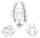
| | | | | | | Ref.: | | | Sewell, 1914 a (p.244, figs.F,M); Steuer, 1923 (p.14, figs.F,M); Sewell, 1924 (p.790, fig.F); 1932 (p.393, Rem.); 1948 (p.324, Rem.); Kasturirangan, 1963 (p.64, figs.F,M); Abraham, 1970 (p.52, Rem.); Silas, 1972 (p.650); Goswami & Goswami, 1978 (p.111, figs.); Madhupratap & Haridas, 1994 (p.67, Redescr., figs.F,M); Barthélémy, 1999 (p.858, 864, figs.F); 1999 a (p.9, Fig.26, A-E); Soh & al., 2013 (p.718, Rem.p.723, Table 1, 2, fig.7, 9: chart). | 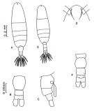 issued from : M. Madhupratap & P. Haridas in Hydrobiologia, 1994, 292/293. [p.69, Fig.1] Female: A, habitus (dorsal view); B, urosome (dorsal), (spinules show variability); C, urosome (right lateral side); D, head with rostral filaments (ventral). Male: F, urosome (dorsal). 20 females and 10 males from the Tuticorin (8°56'N, 78°20'E) and 100 females and 50 males from Portonovo (11°52'N, 79°50'E°E)6), plus specimens from Cochin backwaters (10°N, 76°E), Mandovi-Zuari estuaries (15°30'N, 74°E). Nota female: - Prosome-urosome ratio 3.7 : 1. Cephalosome and pediger 1 separate. -Posterior corners of metasome rounded, naked. - All urosomal somites usually naked. Fine spinules shown on urosomal somites (fig.1B), and male (fig.1F) present only in specimens from Portonovo; the number of spinules varies within this population; specimens examined from other localities without spinules on urosomal somites.; however, spinules may be present on the urosome somites of animals from other localities (Tranter & Abraham, 1971, have shown the female from Cochin possessing some spinules. - Caudal rami symmetrical, 1.4 times longer than broad, bearing 5 plumose setae and 1 setule. Urosomal segments and caudal rami with proportions 46 : 19 : 12: 23 = 100. Nota male: - Prosome-Urosome ratio 3.1 : 1. - Posterior corners of pediger 5 naked. - Urosomal segments also unusually unarmed (variability shown in fig.1F) - Caudal rami nearly as broad as long. - Proportions of urosomal segments and caudal rami 11 : 29 : 23 : 8 : 12 : 17 = 100.
|
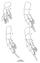
 issued from : M. Madhupratap & P. Haridas in Hydrobiologia, 1994, 292/293. [p.71, Fig.3]. Female: A, A2; B, Md; C, Mx1; E, Mxp. Nota: - A2: coxa with 1 single seta,; basis fused with an endopod segment (allobasis) bearing 8 setae at inner margin, 1 seta terminally and 2 distal spinule rows, 2nd and 3rd segments incompletely fused appearing as a bilobed segment each bearing 7 setae; exopod 4-segmented segments incompletely fused (setal formula: 1, 1, 3, 3). - Md: gnathobase with 5 sharp and 4 blunt teeth; basis with 1 medial seta; endopod with 2 incompletely fused segments, 1st with 2 setae, 2nd with 9 setae; exopod partly fused with basis, 3-segmented (setal formula 1, 1, 4). - Mx1: praecoxal arthrite with 8 spines like and 1 weak setae; coxal endite with 2 setae, coxal epipodite with 9 developed setae; each of 2 basal endites with 1 thick seta, basal exite with 1 seta; endopod and exopod incompletely fused with 5 and 2 setae each. - Mx2: praecoxa and coxa with 2 endites each (setal formula 4, 2, 2, 3); basis with 1 large and 1 small seta; endopod 3-segmented (setal formula 1, 2, 4). - Mxp: praecoxa and coxa fused bearing 5 setae; basis bearing 1 spine like seta; endopod 4-segmented, 1st 3 segments incompletely fused, with 1 seta each; terminal segment with 2 setae; - Mouthparts of male as in female.
|
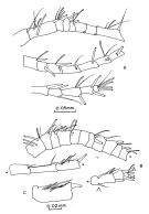
 issued from : M. Madhupratap & P. Haridas in Hydrobiologia, 1994, 292/293. [p.72, Fig.4]. Female: A-D, swimming legs 1-4 (respectively).
|
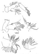
 issued from : M. Madhupratap & P. Haridas in Hydrobiologia, 1994, 292/293. [p.70, Fig.2]. Female: A, A1 (proximal segments, middle segments and distal segments, respectively); Male: right A1 (proximal segments, middle segments and distal segments, respectively); C, segment 14 of right A1. Nota: - A1 female 20-segmented, reaching midlength or pediger 5. Segment 4 with 1 spine, segment 8 longest, segments 6, 7, 11, 12, 14, 15 with spinules, terminal segment with 4 setae and 1 aesthetasc. - Right A1 male 18-segmented, suture between segments 15 and 16 incomplete (arrow head, fig.2B), 2spines present on anterior margin of segment 4, 1 spine each on segments 7 and 12, segments 5 and 6 unarmed, geniculation between segments 13 and 14, segment 14 (fig.2C) armed with 2 larger and 2 smaller spines, 1 seta and 1 aesthetasc.
|
 issued from : M. Madhupratap & P. Haridas in Hydrobiologia, 1994, 292/293. [p.73, Fig.5]. Female: A, P5. Male: B, P5. Nota: - P5 female: symmetrical; 3-segmented, both coxal segments fused; basis oblong with 1 small seta on outer margin at about distal 2/3; base of terminal segment slightly swollen, tapering distally, with a distinct suture at midlength, portion distal to suture serrate. -P5 male: Right and left coxal segments fused. Right leg basis with seta at distolateral corner; 1st exopodal segment with 1 long slender seta, 2nd exopodal segment with an oblong inner lobe bearing 1 spine, 3rd segment curved with 1 thick spine at midlength on medial margin and another at tip. Left leg basis more than twice as long as broad with outer seta at midlength; 1st exopoal segment short and unarmed, terminal segment medial margin with proximal tuft of hair, 1 spine embedded in a membraneous sheath, the sheath produced and of irregular shape and flanked on distal side by a recurved spine; tip of segment bifurcate, outer process much longer than inner process.
|
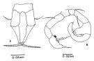
 Issued from : R.B.S. Sewell in Spolia Zeylanica, 1914, 9. [Pl.XIX, Figs.8-9]. Female (from Gulf of Mannar): 9, P5. Nota: Head and 1st thoracic segment separate, 4th and 5th fused. Posterior thoracic margin rounded and devoid of spines. Forehead with a pair of slender curved rostral filaments. Proportional lengths of prosome and urosome 35:1. Proportional lengths of urosomites and furca 26:13:10:15; all segments devoid of any spines. Caudal rami nearly as wide as long (9:10). A1 20-segmented (segments 2-4, 5-6, 7-8, 9-10 fused) reach just beyond the posterior thoracic margin. P5 consist of a fairly long basal segment bearing 1 minute seta on its external margin distally, and having a long and delicate terminal spine, this spine is curved and has a markedly swollen base. Male: 8, P5. Nota: Proportional lengths of prosome and urosome 4:1. Proportional lengths of urosomites and furca 18:14:3:9:10. Right A1 15-segmented (segments 1-4, 5-6, 7-8, 19-21, 22-25 fused) modified to form a grasping organ. P5: the right leg forms a claw, the 2nd segment bears a somewhat quadrangular process on its inner border, and the 3rd segment terminates in 2 short unequal spines.
|
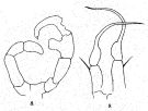
 issued from : R.B.S. Sewell in Fauna of the Chilka Lake, 1924, 12. [Pl. XLV, Fig.6]. Female and Male (from Chilka Lake).
|
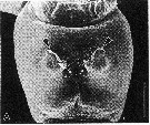 issued from : R.-M. Bathélémy in J. Mar. Biol. Ass. U.K., 1999, 79. [p.864, Fig.7, A]. Scanning electon miccrograph. Female from Indian Ocean): A, genital double-somite (ventral); Note the epicuticule (arrowheads) covering the genital slits except the copulatory field (arrows). Scale bar: 0.020 mm.
|
 Issued from : H.Y. Soh, S.Y. Moon, E.O. Park & B.A.V. Maran in J. Crustacean Biol., 2013, 33 (5). [p.727, Table 1]. Comparison of morphological features between A. forticrusa and its congeners A. southwelli and A. sarojus
| | | | | Compl. Ref.: | | | Subbaraju & Krishnamurphy, 1972 (p.25, 26 ); Patel, 1975 (p.660); Ohtsuka & al., 1995 (p.158: Rem., 159); Madhupratap & Haridas, 1986 (p.105, tab.2); Gajbhiye & Abidi, 1993 (p.137); Godhantaraman, 1994 (tab.5, 6, 7); Mauchline, 1998 (tab.8); Madhu & al., 2007 (p.54, Table 4, abundance vs monsoon); Fernandes, 2008 (p.465, Tabl.2); Rakhesh & al., 2008 (p.154, abundance vs stations); Shanthi & Ramanibai, 2011 (p.132, Table 1); Drillet & al., 2012 (p.155, Table 1, culture); Rakhesh & al., 2013 (p.7, Table 1, abundance vs stations); Varadharajan & Soundarapandian, 2013 (p.2: occurrence vs stations) | | | | NZ: | 2 | | |
|
Distribution map of Acartia (Euacartia) southwelli by geographical zones
|
| | | | | | | | |  issued from : C.T. Achuthankutty, N. Ramaiah & G. Padmavati in Pelagic biogeography ICoPB II. Proc. 2nd Intern. Conf. Final report of SCOR/IOC working group 93, 9-14 July 1995. Workshop Report No. 142, Unesco, 1998. [p.8, Fig.6]. issued from : C.T. Achuthankutty, N. Ramaiah & G. Padmavati in Pelagic biogeography ICoPB II. Proc. 2nd Intern. Conf. Final report of SCOR/IOC working group 93, 9-14 July 1995. Workshop Report No. 142, Unesco, 1998. [p.8, Fig.6].
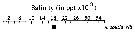
Salinity ranges for A. southwelli in coastal and estuarine waters of Goa (India).
Shaded area indicates the range of higher abundance. |
| | | | Loc: | | | India ( Saurashtra coast, S, Cochin, Kerala, Porto Novo, G. of Mannar, Sri Lanka Pearl Banks, Pointcalimere-Mallipattinam, Godavari estuary & coastal, Kakinada Bay, Chilka Lake), Bay of Bengal, China Seas (Bohai Sea) | | | | N: | 19 | | | | Lg.: | | | (44) F: 0,773; M: 0,68; (80) F: 0,726; M: 0,712; (82) F: 0,8; M: 0,75; (186) F: 0,84-0,79; M: 0,75-0,7; {F: 0,726-0,84; M: 0,68-0,75} | | | | Rem.: | estuary-neritic. Brackish.
After Madhupratap & Haridas (1994), the female described from Chilka Lake by Sewell (1924) from a smaller form (0.726 mm) seems to be based on an immature specimen (stage copepodite V).
The dimensions seem to be correlated with the salinity.
W. Zhang confirms the occurence of this species in the China seas (in the Bohai Sea, 2010).
For Ohtsuka & al. (1995, p.158) A. southwelli, as Bestiolina similis and Pseudodiaptomus marinus belong to the Indo-West Pacific warm-water element which is thought to have derived directly from the Tethys fauna (see in Nishimura, 1981). These copepods seem prefer relatively high salinities. | | | Last update : 06/06/2020 | |
|
|
 Any use of this site for a publication will be mentioned with the following reference : Any use of this site for a publication will be mentioned with the following reference :
Razouls C., Desreumaux N., Kouwenberg J. and de Bovée F., 2005-2026. - Biodiversity of Marine Planktonic Copepods (morphology, geographical distribution and biological data). Sorbonne University, CNRS. Available at http://copepodes.obs-banyuls.fr/en [Accessed June 09, 2026] © copyright 2005-2026 Sorbonne University, CNRS
|
|
|
|


 southwelli - Plate 1 of morphological figures',750,500);)
 southwelli - Plate 2 of morphological figures',750,500);)
 southwelli - Plate 3 of morphological figures',750,500);)
 southwelli - Plate 4 of morphological figures',750,500);)
 southwelli - Plate 5 of morphological figures',750,500);)
 southwelli - Plate 6 of morphological figures',750,500);)
 southwelli - Plate 7 of morphological figures',750,500);)
 southwelli - Plate 8 of morphological figures',750,500);)

 southwelli - Plate 1 of morphological figures)
 southwelli - Plate 2 of morphological figures)
 southwelli - Plate 3 of morphological figures)
 southwelli - Plate 4 of morphological figures)
 southwelli - Plate 5 of morphological figures)
 southwelli - Plate 6 of morphological figures)
 southwelli - Plate 7 of morphological figures)
 southwelli - Plate 8 of morphological figures)
 southwelli - Plate 9 of morphological figures)
 southwelli - Distribution map 2)
