|
|
 |
|
Calanoida ( Order ) |
|
|
|
Arietelloidea ( Superfamily ) |
|
|
|
Augaptilidae ( Family ) |
|
|
|
Euaugaptilus ( Genus ) |
|
|
| |
Euaugaptilus magnus (Wolfenden, 1904) (F,M) | |
| | | | | | | Syn.: | Augaptilus magnus Wolfenden, 1904 (p.122, Descr.F); Pearson, 1906 (p.28); Farran, 1908 b (p.77, Rem.); Wolfenden, 1911 (p.337, 341, figs.F);
Augaptilus fungiferus Steuer, 1904 (p.597); no Wolfenden, 1911 (p.336, figs.F);
? Augaptilus validus A. Scott, 1909 (p.138, figs.F,M);
E. magnus fungiferus : Sewell, 1947 (p.213-214, figs.F, Rem.);
E. magnus magnus : Vervoort, 1965 (p.139, Rem.);
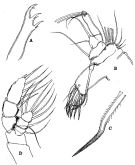
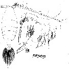
Euaugaptilus squamatus : Lysholm & al., 1945 (part., p.38, 39, 48) | | | | Ref.: | | | Sars, 1925 (p.262, figs.F,M); Farran, 1926 (p.289); Rose, 1929 (p.35); Sewell, 1932 (p.322); Rose, 1933 a (p.222, figs.F,M); Jespersen, 1934 (p.112); 1940 (p.58); Sewell, 1947 (p.213, figs.F, Rem.); ? Vervoort, 1957 (p.139, Rem.); Tanaka, 1964 b (p.53, figs.F,M); Tanaka & Omori, 1967 (p.253); Owre & Foyo, 1967 (p.87, figs.F,M) ; Matthews, 1972 (p.23: body length variation, p.35: Rem.); Tanaka & Omori, 1974 (p.235, figs.M); Roe, 1984 (p.358); Park, 1993 (p.41, figs.F, M, Rem.); Bradford-Grieve & al., 1999 (p.882, 939, figs.F,M) ; Bradford-Grieve,1999 b (p.55, Rem., figs.172, 190) Matsuura & Nishida, 2000 (p.339); Vives & Shmeleva, 2007 (p.201, figs.F,M, Rem.) | 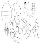 issued from : O. Tanaka in Publs Seto Mar. Biol. Lab., 1964, XII (1). [p.54, Fig.199]. Female: a, habitus (dorsal); b, forehead (left lateral side); c, last thoracic segment and urosome (left lateral sifde); d, Md (mandibular palp and bitting blade); e, exopod of P1; f, P5. Nota: The urosome segments and furca are in the proportional lengths as 55:15:9:21 = 100. Male: g, P5 (with other view of the distal segment of exopod of left leg). Nota: The urosome segments and furca are in the proportional lengths as 26:17:15:11:9:22 = 100. The mouth parts and swimming legs as in the female; the right A1 extends to the end of the furca; the grasping on the left A1.
|
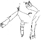
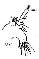
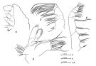 issued from : O. Tanaka & M. Omori in Publs Seto Mar. Biol. Lab., 1974, XXI (3/4). [p.236, Fig.19]. Male: a, head (lateral); b, Mx1; c, Mx2; d, Mxp.
|
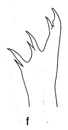 issued from : O. Tanaka & M. Omori in Publs Seto Mar. Biol. Lab., 1974, XXI (3/4). [p.199, Fig.2,f]. Female: f, mandibular teeth.
|
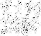
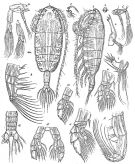 Issued from : G.O. Sars in Résult. Camp. Scient. Prince Albert I, 69, pls.1-127 (1924). [Pl.LXXIX, figs.1-16]. Female: 1, habitus (dorsal); 2, idem (lateral left side); 3, forehead (lateral); 4, rostral prominence; 5, A2; 6, Md; 7, Mx1; 8, Mx2; 9, Mxp; 10, P1; 11, P2; 12, P5. Male: 13, A2; 14, Md (mandibular palp); 15, P5; 16, urosome (dorsal).
|
 issued from : R.B.S. Sewell in The John Murray Expedition, 1933-34, Scientific Reports, VIII (1), 1947. [p.215, Fig.57]. As Euaugaptilus magnus forma fungiferus. Female (from Arabian Sea and G. of Aden): A, Md (biting blade-; B, Mx1; C, One of the spines of the inner lobe (enlarged); D, P5. Nota: Proportional lengths of the prosome and urosome as 76 to 24. The proportional lengths of the various segments of the body (cephalon to caiudal rami) as 414:130:65:65:85:129:34:24:44 = 1000. The arrangement and number of setae arising from the different parts of Mx1 agree with the accounts of both magnus and fungiferus
|
 issued from : T. Park in Biology of the Antarctic Seas, XXII. Antarct. Res. Ser., Washington, 58. [p.42, Fig.29]. Female: a, forehead (lateral); b, A2; c, Md; d, Md (biting edge); e, Mx1; f, Mx2.
|
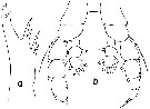 issued from : T. Park in Biology of the Antarctic Seas, XXII. Antarct. Res. Ser., Washington, 58. [p.43, Fig.30]. Male: a, Md (biting edge); b, P5 (posterior).
|
 issued from : R.N. Wolfenden in Die Marinen Copepoden der Deutschen Südpolar-Expedition 1901-1903, 1911. [p.342, Fig.74]. As Augaptilus magnus. Female: a, A2; b, Md.
|
 issued from : R.N. Wolfenden in Die Marinen Copepoden der Deutschen Südpolar-Expedition 1901-1903, 1911. [Pl.XXXVII, Figs.4-9]. As Augaptilus magnus. Female: 4, habitus (dorsal); 5, Mx1; 6, Mxp; 7, structure of setal portion of Mxp; 8, P1; 9, P5.
|
issued from : R.N. Wolfenden in Die Marinen Copepoden der Deutschen Südpolar-Expedition 1901-1903, 1911. [Pl.XXXVII, Figs.4-9]. As Augaptilus magnus. Female: 4, habitus (dorsal); 5, Mx1; 6, Mxp; 7, structure of setal portion of Mxp; 8, P1; 9, P5.
|
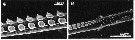 issued from : H. Matsuura & S. Nishida in Mar. Biol., 2000, 137. [p.341, Fig.2, C-D]. Female (from eastern Indian and NW Pacific): SEM micrographs of button setae on Mx2 and Mxp: C, subterminal part of a seta; D, transitional part of a seta. Nota: Observe the semicircular disc shape. Number of button setae on Mx2: 1 on basis, 9 on endopod; on Mxp: 7 on endopod.
|
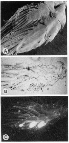 issued from : G.A. Boxshall in Microscopic Anatomy of Invertebrates. 9: Crustacea. 1992. Wiley-Liss, Inc. [p.356, Fig.6, A-C]. A: P3 showing pores of bioluminescent glands (arrowed). B: Lightmicrograph of P3 showing 3 sets of bioluminescent glands (one set arrowed). G: Fluorescence micrograph of B.
|
 issued from : J.M. Bradford-Grieve, E.L. Markhaseva & C.E.F. Rocha & B. Abiahy in South Atlantic Zooplankton, Edit. D. Boltovskoy. 1999. Vol.2. Copepoda. [p.1039, Fig. 7.255]. Female (S Atlantic): Mx1. Nota: Exopod with 3 setae; outer lobe 1 with 8 setae.
|
 issued from : J.B.L. Matthews in Bull Gr. Mus. (Nat. Hist.) Zool., 1972, 24 (1). [p.25, Table I). Occurrence of a seta on the posterior surface near the outer margin of basipodite of P1, P4 and P5. Numbers refer to the number of individuals examined. Sars (1925) figured Mx1 with 10 setae on the 1st endite, 1 on the 2nd, 1 small one on the 3rd; 1 on the basipodite, 2 on the exopodite; and 8 on the exite. Matthews (1972, p.24), in the specimens examined found invariably 11 on the 1st endite. The 2nd endite was also constant, with 1 seta, but on the 3rd endite rhe single seta was further reduced in some specimens and in one case was absent altogether. The basipodite of one appendage out of the 28 examined bore a small additional seta. The exopodite in all cases had two principal setae, as described, but in most cases there were 1, 2 or 3 subsidiary ones, so small as easily to be overlooked. The setation of the exite also showed some variation in the number of minor setae at the proximal end of the row.
| | | | | Compl. Ref.: | | | Sewell, 1948 (p.330, 504, 522, 531, 534, 539, 547); C.B. Wilson, 1950 (p.206); Grice, 1963 a (p.496); De Decker & Mombeck, 1964 (p.12); Grice & Hulsemann, 1967 (p.18); 1968 (tab.2); Itoh, 1970 (tab.1); Park, 1970 (p.478); Morris R.J., 1971 (p.275, Table 1, 2, lipids composition); Roe, 1972 (p.277, tabl.1, tabl.2); Macdonald & al., 1972 (p.213, fig.4, hydrostatic pressure effect); Björnberg, 1973 (p.348, 385); Harding, 1974 (p.141, tab. 2, gut contents); Vives & al., 1975 (p.50, tab.II, XII); Deevey & Brooks, 1977 (p.156, tab.2, Station "S"); Vives, 1982 (p.294); Guangshan & Honglin, 1984 (p.118, tab.); Lozano Soldevilla & al., 1988 (p.60); Lapernat, 2000 (tabl.3, 4); Razouls & al., 2000 (p.343, Appendix); d'Elbée, 2001 (tabl.1); Holmes, 2001 (p.12); Hsiao & al., 2004 (p.325, tab.1); Fernandes, 2008 (p.465, Tabl.2); Gaard & al., 2008 (p.59, Table 1, N Mid-Atlantic Ridge); Park & Ferrari, 2009 (p.143, Table 4, Appendix 1, biogeography); Hidalgo & al., 2010 (p.2089, Table 2); Matsuura & al., 2010 (p.2098, Table 2, 3, fig.8) | | | | NZ: | 16 + 1 doubtful | | |
|
Distribution map of Euaugaptilus magnus by geographical zones
|
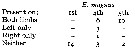
| | | | | | | | | | | | | | | 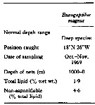 Issued from : R. J. Morris in Comp. Biochem. Physiol., 1971, 40B. [p.277, Table 1]. Issued from : R. J. Morris in Comp. Biochem. Physiol., 1971, 40B. [p.277, Table 1].
Total lipid and saponification for Euaugaptilus magnus from Northeastern Atlantic. |
| | | | Loc: | | | Antarct. (SW Atlant., Indien, SW & SE Pacif.), sub-Antarct. (Indien, SW & SE Pacif.), South Africa (E), off St. N Helena, G. of Guinea, off NE St. Paul Is., Cape Verde Is., off NW Cape Verde Is., Canary Is., off Canaries W, off Madère, Mer des Antilles, Floride, Mer des Sargasses, off Bermuda 'Station "S"), off E Cape Cod, Labrador, S Strait of Davis, S Grrenland, Iceland, off W Irrland, Bau of Biscay, off W Cape Finisterre, Ibero-moroccan Bay, ? Medititerranean Sea, G. of Aden, Arabian Sea, Indian, Is. Laccadive Is., Bay of Bengal, Indonesia-Malaysia, Celebes and Sulu Seas, China Seas (South China Sea), E Taiwan , Japan (Izu), Sea of Philippines, Pacif. (W equatorial), Tasman Sea, SW Galapagos, off Peru, off Juan Fernandez Is., Chile (N & S) | | | | N: | 44 | | | | Lg.: | | | (1) F: 8,4; (10) F: 6,65; (38) F: 6,96; M: 6,96; (48) F: 8,58-7,33; M: 8,25-8,08; (68) F: 8,9-6,58; M: 8,5-8,1; (69) F: 7,92; M: 7,75; (70) F: 8,6-7,5; M: 8,8-8,7; (199) F: 8-7,02; (1066) F: 7,3-7,7; {F: 6,58-8,90; M: 6,96-8,80} | | | | Rem.: | meso-bathy-abyssopelagic. Obtained at depths between 440 and 2500 m, occasionally near the surface (Matthews (1972, p.36)
Sampling depth (Antarct., sub-Antarct.): 1500 m. Sargasso Sea: 500-2000 m (Deevey & Brooks, 1977, Station "S"); 2000-1000 m (Harding, 1974).
The specimens from the Izu region showed a considerable variability in the body length (6.58 to 8.90 mm); the two forms E. magnus fungiferus (Sewell, 1947) and E. magnus magnus are present.
For Park (1993, p.44) this species is closely related to E. austrinus, but can be distinguished from it, Particulary by the shape of Md: the mandibular palp is relatively small, the toothed edge of the mandibular blade is strongly oblique to the long axis of the blade, and the teeth are relatively long in contrast to E. austrinus. Meanwhile the number of setae on A2, Md, Mx1 and Mx2, however, show a small degree of variation. Of the 42 females examined, the number of specimens that show an aberrant setal arrangement is as follows: the 1st endopodal segment of A2 had no seta, instead of having 1, in two specimens. 1st endopodal segment of mandibular palp had no seta, instead having 1, in two specimens. 3rd inner lobe of Mx1 had no seta, instead of having 1, in 4 specimens. Basis of Mx1 had 2 setae, instead of 1, in 4 specimens. Exopod of Mx1 had 3 long setae, instead of 2, in 2 specimens. 1st protopodal lobe of Mx2 had 2 setae, instead of 3, in 2 specimens.
Farran (1908) suggested that Augaptilus fungiferus Steuer, 1904, from the Indian Ocean is a synonym of E. magnus. Sewell (1947), on the other hand, considered A. fungiferus and A. validus Scott, 1909 (from the Malay Archipelago), synonymous and then regarded them as a form of E. magnus. According to Sewell's 1947 descriptions based on specimens from the Arabian Sea, Euaugaptilus f fungiferus is smaller (body length = 7.08-7.41) than E. magnus found by Park (body length 8.58-7.33), but otherwise seems to agree in anatomical details with the latter. Park (1993) follow Matthews' s 1972 opinion that A. fungiferus Steuer and A. validus Scott are synonyms of E. magnus.
For Itoh (1970 a, fig.2, from co-ordonates) the Itoh's index value from mandibular gnathobase = 1550.
For Matthews (1972, p.23) the Md gnathobase show 6 well-defined teeth, the number of teeth was the same in all specimens examined and there was no noticeable difference in their arrangement.
In ''squamatus" Group after Matthews (1972, p.64) {See remarks in Euaugaptilus genus}. | | | Last update : 04/12/2020 | |
|
|
 Any use of this site for a publication will be mentioned with the following reference : Any use of this site for a publication will be mentioned with the following reference :
Razouls C., Desreumaux N., Kouwenberg J. and de Bovée F., 2005-2025. - Biodiversity of Marine Planktonic Copepods (morphology, geographical distribution and biological data). Sorbonne University, CNRS. Available at http://copepodes.obs-banyuls.fr/en [Accessed December 01, 2025] © copyright 2005-2025 Sorbonne University, CNRS
|
|
|
|


;)
;)
;)
;)
;)
;)
;)
;)
;)
;)
;)
;)

;)
