|
|
 |
|
Calanoida ( Order ) |
|
|
|
Clausocalanoidea ( Superfamily ) |
|
|
|
Clausocalanidae ( Family ) |
|
|
|
Microcalanus ( Genus ) |
|
|
| |
Microcalanus pygmaeus (Sars, 1900) (F,M) | |
| | | | | | | Syn.: | Pseudocalanus pygmaeus Sars, 1900 (p.73, Descr.F, figs.F); Giesbrecht, 1902 (p.20, figs.F); Mrazek, 1902 (p.508, figs.F,M);
Spinocalanus longicornis (M) Sars, 1900 (p.77, figs.M);
? Microcalanus pusillus Sars, 1903 (p.156, figs.F,M);
Microcalanus pygmaeus pygmaeus : Bradford-Grieve, 1994 (p.152, fig.96); Dvoretsky & Dvoretsky, 2010 (p.991, Table 2);
Microcalanus pygmaeus pusilus : Wishner & al., 2008 (p.163, Table 2, fig.8, oxycline) | | | | Ref.: | | | Sars, 1901 a (1903) (p.20, 156); With, 1915 (p.66, Rem.); Farran, 1926 (p.242, Rem.); 1929 (p.208, 226); Rose, 1933 a (p.80, figs.F,M); Jespersen, 1934 (p.50); 1940 (p.15); Wilson, 1942 a (p.194, fig.M); Brodsky, 1950 (1967) (part., p.116, figs.F,M); Farran & Vervoort, 1951 e (n°37, p.3, figs.F); Wiborg, 1954 (p.141, Rem.); 1955 (p.38, Rem.); Tanaka, 1956 c (p.385, figs.F, Rem.); Vervoort, 1957 (p.36, Rem.); Tanaka, 1960 (part., p.35, figs.F,M, juv.M, Rem.); Fagetti, 1962 (p.15); Minoda, 1971 (p.19); Vidal, 1971 a (p.17, 24, figs.F,M, Rem., p.116); Bradford, 1971 b (p.16, figs.F,M, Rem.); Kos, 1976 (Vol.II, figs.F,M, Rem.); Björnberg & al., 1981 (p.628, figs.F,M); Brodsky & al., 1983 (p.226, figs.F,M); Mazzocchi & al., 1995 (p.171, figs.F,M, Rem.); Menshenina & Melnikov, 1995 (p.128); Chihara & Murano, 1997 (p.779, Pl.95: F,M); G. Harding, 2004 (p.46, figs.F,M); Michels & Schnack-Schiel, 2005 (p.483, fig.7: Md); Conway, 2006 (p.11, 25, copepodite stages 1-6, Rem.); Vives & Shmeleva, 2007 (p.634, figs.F,M, Rem.) | 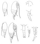 Female: 1, habitus (dorsal view); Issued from : Tanaka O. in Publs Seto mar. Biol. Lab., 1956, 5 (3): 367-406. 2, habitus (lateral); 4, Urosome (lateral); issued from Vidal, 1971; 3, Head (lateral); issued from Tanaka, 1960. Male: 5, habitus; 7, Urosome (dorsal); 8, P5; issued from : Brodsky, Vyshkvartseva, Kos & Markhaseva in Opred. Fauna SSSR, 1983, 135: 1-357. 6, habitus (lateral); 9, P5; issued from Vidal, 1971
|
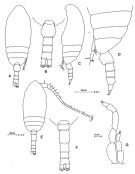 Issued from: M.G. Mazzocchi, G. Zagami, A. Ianora, L. Guglielmo & J. Hure in Atlas of Marine Zooplankton Straits of Magellan. Copepods. L. Guglielmo & A. Ianora (Eds.), 1995. [p.172, Fig.3.30.1]. Female: A, habitus (dorsal); B, urosome (dorsal); C, habitus (lateral left side); D, 3rd-5th thoracic somites and urosome (lateral left side). Nota: A1 reaching half genital somite. Proportional lengths of urosomites and furca 33:19:16:17:15 = 100. Male: E, habitus (dorsal); F, urosome (dorsal); G, P5. Nota: Proportional lengths of urosomites and furca 12:30:20:14:13:11 = 100.
|
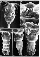 Issued from: M.G. Mazzocchi, G. Zagami, A. Ianora, L. Guglielmo & J. Hure in Atlas of Marine Zooplankton Straits of Magellan. Copepods. L. Guglielmo & A. Ianora (Eds.), 1995. [p.173, Fig.3.30.2]. Female (SEM preparation): A, habitus (dorsal); B, forehead (ventral); C, urosome (dorsal); D, idem (lateral right side); E, idem (ventral). Bars: A 0.100 mm; B, C, D, E 0.010 mm.
|
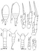 issued from : K.A. Brodsky, N.V. Vyshkvartzeva, M.S. Kos & E.L. Markhaseva in Opred Fauna SSSR, 1983, 135. [p.226, Fig.106]. Female & Male (from Okhotsk Sea).
|
 issued from : J. Michels & S.B. Schnack-Schiel in Mar. Biol., 2005, 146. [p.489, Fig.7]. Mandibular gnathobase. a: Female (from Weddell and Bellingshausen Seas); b, Male. a, b,: left gnathobase from cranial; V: ventral tooth, C1-C4: central teeth, D1-D3: dorsal teeth, B: dorsal bristle; arrow: the male gnathobase.. Scale bars 0.020 mm. Nota: This species seems to feed mainly on phytoplankton (Hopkins & Torres, 1989; Pasternak & Schnack-Schiel, 2001), however, the gnathobase morphology, with very long and pointed teeth that are not suitable for feeding on phytoplankton, points to another feeding behaviour. The gnathobase teeth are very similar to those of Paraeuchaeta antarctica. It must beassumed that M. pygmaeus also feeds on zooplankton.. The mandibular gnathobases possess two teeth more than those of P. antarctica, which permits the classification of M. pygmaeus as a phyto-and zooplankton feeder, confirmed by the results of Hopkins, 1985 and Hopkins & al., 1995, who found pieces of metazoans in the guts of M. pygmaeus. This species not only feeds on phytoplankton, but also on zooplankton and detritus in deep water layers during winter and early spring.
|
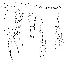 issued from : W. Giesbrecht in Copepoden. Res. voyage du S. Y. Belgica. Rapports scientifiques, Zoologie, 1902. [Taf. II, Figs. 1-5]. As Pseudocalanus pygmaeus. Female (from Antarctic): 1, habitus (lateral); 2, A1; 3, urosome (dorsal); 4, P2; 5, P4.
|
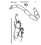 Issued from : J.M. Bradford in N.Z. Oceanogr. Inst., 1971, 206, Part 8, No 59. [p.15, Figs.10, 11]. Male (from 71°15'S - 75°55'S, 172°20'E - 175°22'W): 10, P5. Female: 11, P1.
|
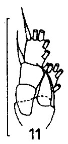 Issued from : J.M. Bradford in N.Z. Oceanogr. Inst., 1971, 206, Part 8, No 59. [p.15, Fig. 11]. Female (from Ross Sea): 11, P1.
|
 Issued from : J.M. Bradford in N.Z. Oceanogr. Inst., 1971, 206, Part 8, No 59. [p.15, Fig.10]. Male (from 71°15'S - 75°55'S, 172°20'E - 175°22'W): 10, P5.
|
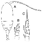 issued from : J.M. Bradford-Grieve in The Marine Fauna of New Zealand: Pelagic Calanoid Copepoda. National Institute of Water and Atmospheric Research (NIWA). New Zealand Oceanographic Institute Memoir, 102, 1994. [p.130, Fig.75, A-C]. From Sars, 1900. As Microcalanus pygmaeus pygmaeus. Female (from 62°S, 170°E): A, habitus (dorsal); B, P1; C, P2.
|
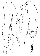 Issued from : O. Tanaka in Spec. Publs. Seto mar. biol. Lab., 10, 1960 [Pl. XIII, 1-8]. Female (from 67°S, 40°50'E): 1, forehead (lateral); 2, P2; 3, P3; 4, forehead (lateral) other specimen. Male: 5, habitus (dorsal); 6, forehead (lateral); 7, last thoracic segment with P5 and urosome (lateral); 8, P5. Nota: The Tanaka's antarctic specimens belong to the sub-species M. pygmaeus pygmaeus in having the long A1. Two size groups were found among specimens: the small form measured 0.60-0,74 mm, and the large dform measured 0,83 mm. The small form has rather short rostral spines, whereas, the large form has long rostral spines. The terminal spine of the 3rd segment of P2 is rather coarsely dentated (about 40 teeth) in the small form, whereas, it is more finely dentated (about 50) in the latter. The body is transparent in the large form.
|
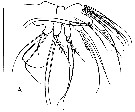 Issued from : E.L. Markhaseva & J. Renz in Crustaceana, 2015, 88 (9). [p.1043, Fig.6, A]. Female: A, Mx2. Scale bar: 0.1 mm.
|
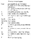 Issued from : C. Razouls in Ann. Inst. océanogr., Paris, 1994, 70 (1). [p.42]. Caractéristiques morphologiques de Microcalanus pygmaeus femelle et mâle adultes. Terminologie et abbréviations: voir à Calanus propinquus. Nota: Peut être confondu avec Microcalanus pusillus Sars, 1903 qui se distingue de la précédente par A1 (female et mâle) qui ne dépasse pas le segment génital, les segment distaux (Exo 3) de P3, P4 et P5 moins élancés, la soie terminale à dents plus grossières; la P5 gauche du mâle égale au tiers de la droite.
|
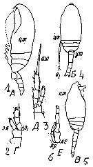 Issued from : M.S. Kos in Field guide for plankton. Zool Institute USSR Acad., Vol. II, 1976. After Brodsky, 1950. Female: 1, habitus (dorsal); 2, P1; 3, P3. Male: 4-5, habitus (dorsal and lateral, respectively); 6, P5 (np = right, le = left)
|
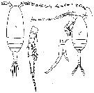 Issued from : M. Rose in Faune de France, 1933, 26. [p.80, Fig. 35]. Female: habitus (dorsal), and P3. Male: habitus (dorsal) and P5. Nota: Different pseudoàcalanus by exopod al segment 1 of P1 without outer spine and endopod of P1 with 4 setae. Left P5 slernder, s (or ? 6 segments) distal segment of right P5 short, not stylet-like. Female: Length A1 less than the end of the caudal rami. Male: right P5 two times less long than long than the left.
| | | | | Compl. Ref.: | | | Damas & Koefoed, 1907 (p.396, tab.II); Hardy & Gunther, 1935 (1936) (p.149, Rem.); Jespersen, 1939 (p.44, Rem., Table 25, 26, 27, 28, 29, 30); Lysholm & al., 1945 (p.11); Sewell, 1948 (p.395, 512, 453, 487, 513); Gundersen, 1953 (p.1, 18, Table 17, seasonal abundance); Østvedt, 1955 (p.14: Table 3, p.17, 57: Rem.); Minoda, 1958 (p.253, Table 1, 2, abundance); Grice, 1962 a (p.101); M.W. Johnson, 1963 (p.89, Table 1, 2); Unterüberbacher, 1964 (p.19); De Decker & Mombeck, 1964 (p.13); Grice & Hulsemann, 1965 (p.223); Furuhashi, 1966 a (p.295, vertical distribution vs mixing Oyashio/Kuroshio region); Harding, 1966 (p.17, 65); Grice & Hulsemann, 1967 (p.14); Maclellan D.C., 1967 (p.101, 102: occurrence); Vinogradov, 1968 (1970) (p.256, 266); Park, 1970 (p.475); Shih & al., 1971 (p.44, 150, 206); McLaren, 1978 (p.1330, 1335: life history); Kosobokova, 1980 (p.167, seasonal changes, vertical distribution, age composition); Gagnon & Lacroix, 1981 (p.401, Table 1, tidal effect); Buchanan & Sekerak, 1982 (p.41, Rem.: p.47); Brenning, 1982 a (p.11, spatial distribution & T-S diagram, Rem.); Kovalev & Schmeleva, 1982 (p.83); Huntley & al., 1983 (p.143, Table 2); De Ladurantaye & al., 1984 (p.21, tab.I, fig.3, 4, 5, advective processes in fjord); Sameoto, 1984 (p.213, Table 1); Tremblay & Anderson, 1984 (p.6); Hopkins, 1985 (p.197, Table 1, gut contents); Schnack & al., 1985 (p.256, fig.4); Brenning, 1985 a (p.16, 28, Table 2); Groendahl & Hernroth, 1986 (tab.1); Zmijewska, 1987 (tab.2a); Hopkins & Torres, 1988 (tab.1); Ward, 1989 (tab.2); Perissinotto, 1989 (p.505, Table 2, 3, abundance, diurnal variation); Kosobokova, 1989 (p.27); Anderson J.T., 1990 (p.127, Rem.: p.131); Øresland, 1990 (p.201, Table 1, predator); Kellermann, 1990 (p.159, Table 1, predation); Shih & Marhue, 1991 (tab.2, 3); Hattori, 1991 (tab.1, Appendix); Norrbin, 1991 (p.421, life history, gonad maturation); Menshenina & Rakuza-Suszczewski, 1992 (1993) (p.65); Mumm, 1993 (tab.1, fig.2); Kurbjeweit & al., 1993 (p.255, fig.2); Vinogradov & al., 1994 (tab.1); Richter, 1994 (tab.4.1a); Schnack-Schiel & Mizdalski, 1994 (p.357, seasonal variations); Petryashov & al., 1995 (tab.1); Ward & al., 1995 (p.195, abundance, biomass, vertical distribution); Abramova, 1996 (tab.1); Knox & al., 1996 (tab.1); Errhif & al., 1997 (p.422); Park & Choi, 1997 (Appendix); Atkinson, 1998 (p.289, Table 1, biological data); Elwers & Dahms, 1998 (p.150, 151); Kosobokova & al., 1998 (tab.2); Mauchline, 1998 (tab.47, 58); Dolganova & al., 1999 (p.13, tab.1); Abramova, 1999 (p.161, Table 2); Voronina & Kolosova, 1999 (p.71); Goldblatt & al., 1999 (p.2619, tabl. 2); Harvey & al., 1999 (p.1, 49: Appendix 5, in ballast water vessel); Bragina, 1999 (p.195); Atkinson & Sinclair, 2000 (p.46, 50, 51, 53, 55, zonal distribution); Razouls & al., 2000 (p.343, tab. 5, Appendix); Pinchuk & Paul, 2000 (p.4, table 1, % occurrence); Kosobokova & Hirche, 2000 (p.2029, tab.2); Musaeva & Gagarin, 2000 (p.534, tab.1); Zmijewska & al., 2000 (p.89); Musaeva & Suntsov, 2001 (p.511); Fortier M. & al., 2001 (p.1263, fig.6, 7, diel vertical migration); Chiba & al., 2001 (p.95, tab.4, 7); Pasternak Schnack-Schiel, 2001 (p.25); Yamaguchi & al., 2002 (p.1007, tab.1); Auel & Hagen, 2002 (p.1013, tab.2, 3); Ward & al., 2002 (p.2183, tab.2); Ringuette & al., 2002 (p.5081, Table 1, Fig.7, population dynamic); Cabal & al., 2002 (p.869, Table 1, abundance); Ward & al., 2003 (p.121, tab.4); Ashjian & al., 2003 (p.1235, figs.); Hunt, 2004 (p.1, 74, Table 4.4, fig 4.7); Park, W & al., 2004 (p.464, tab.1); Hopcroft & al., 2005 (p.198, table 2); Shimode & al., 2005 (p.113 + poster); Ward & al., 2005 (p.421, Table 6, abundance); Ward & al., 2006 (p.83: tab.4); Tsujimoto & al., 2006 (p.140, Table1); Hop & al., 2006 (p.182, Table 4); Deibel & Daly; 2007 (p.271, Table 1, 2, 4, 6b, Rem.: Arctic, Antarctic polynyas); Ward & al., 2007 (p.1871, Table 2, fig.6, abundance); Kattner & al., 2007 (p.1628, Table 1); Schnack-Schiel & al., 2008 (p.1045: Tab.2, p.1048: fig.5); Ward & al., 2008 (p.241, Tabls, Appendix II ); Humphrey, 2008 (p.84: Appendix A); Darnis & al., 2008 (p.994, Table 1, figs.8, 9); Galbraith, 2009 (pers. comm.); Dvoretsky & Dvoretsky, 2009 a (p.11, Table 2, abundance); Park & Ferrari, 2009 (p.143, Table 4, Appendix 1, biogeography); C.E. Morales & al., 2010 (p.158, Table 1); Eloire & al., 2010 (p.657, Table II, temporal variability); Homma & Yamaguchi, 2010 (p.965, Table 2); Schnack-Schiel & al., 2010 (p.2064, Table 2: E Atlantic subtropical/tropical); Hidalgo & al., 2010 (p.2089, fig.4, Table 2, cluster analysis); Hopcroft & al., 2010 (p.27, Table 2, fig.9); Mazzocchi & Di Capua, 2010 (p.425); Medellin-Mora & Navas S., 2010 (p.265, Tab. 2); Swadling & al., 2010 (p.887, Table 2, 3, A1, fig.6, abundance, indicator species); Kosobokova & al., 2011 (p.29, Table 2, Rem.: Arctic Basins); Pomerleau & al., 2011 (p.1779, Table III); Dvoretsky & Dvoretsky, 2011 a (p.1231, Table 2: abundance, biomass); Homma & al., 2011 (p.29, Table 2, 3, 4, abundance, feeding pattern: suspension feeders); Matsuno & al., 2011 (p.1349, Table 1, abundance vs years); Matsuno & al., 2012 (Table 2); Ward & al., 2012 (p.78, Table A1, B1, abundance, weight); DiBacco & al., 2012 (p.483, Table S1, ballast water transport); Dvoretsky & Dvoretsky, 2012 (p.1321, Table 2, 3, 4, 5, abundance, biomass, production); Makabe & al., 2012 (p.432, Table II, abundance vs mesh-size used); Michels & al., 2012 (p.369, Table 1, fig.8, occurrence frequency, vertical distribution); Hidalgo & al., 2012 (p.134, Table 2); Ward & al., 2014 (p.305, Table 4, 6, 7, seasonal and abundance in the ''Discovery'' Investigations in the 1930s); Ohashi & al., 2013 (p.44, Table 1, Rem.); Ojima & al., 2013 (p.1293, Table 2, 3, 4, fig.3, abundance); Lee D.B. & al., 2013 (p.1215, Table 1, abundance, composition); Dvoretsky & Dvoretsky, 2013 a (p.205, Table 2, % abundance); Eisner & al., 2013 (p.87, Table 3: abundance vs water structure); Pino-Pinuer & al., 2014 (p.83, Table 1, 2, fig.3, abundance variation vs time); Coyle & al., 2014 (p.97, table 3); Smoot & Hopcroft, 2016 (p.1, Rem.: p.7, fig.3, abundance vs water mass); Benedetti & al., 2016 (p.159, Table I, fig.1, functional characters); Marques-Rojas & Zoppi de Roa, 2017 (p.495, Table 1); Belmonte, 2018 (p.273, Table I: Italian zones) | | | | NZ: | 18 | | |
|
Distribution map of Microcalanus pygmaeus by geographical zones
|
| | | | | | | | | | | | | | | 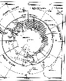 issued from : W. Vervoort in B.A.N.Z. Antarctic Reseach Expedition, Reports - Ser. B, Vol. III, 1957 [Fig.9]. issued from : W. Vervoort in B.A.N.Z. Antarctic Reseach Expedition, Reports - Ser. B, Vol. III, 1957 [Fig.9].
Chart showing the geographical distribution (white circle) in the seas surrounding the Antarctic continent.
Nota: In this chart the area frequented by whaling vessels has been hatched. The Antarctic circle (66°.5 S) has been drawn as a broken line. The numbers I to VI refer to the sectors into which the Antarctic seas are divided according to Mackintosh (1942) (after Vervoort, 1951). |
 issued from : U. Brenning in Wiss. Z. Wilhelm-Pieck-Univ. Rostock - 31. Jahrgang 1982 a. Mat.-nat. wiss. Reihe, 6. [p.11, Figs.1, 2]. issued from : U. Brenning in Wiss. Z. Wilhelm-Pieck-Univ. Rostock - 31. Jahrgang 1982 a. Mat.-nat. wiss. Reihe, 6. [p.11, Figs.1, 2].
Spatial distribution and T-S diagram for Microcalanus pygmaeus from 8° S - 26° N; 16°- 20° W, for expedition V1: Dec. 1972- Jan. 1973.
SO: Southern Surface Water (S °/oo: 34,50; T°C: 29,0); ND: Northern Water of the Surface Layer (S °/oo: 37,5; T°C: 21,0); SD: Southern Deep Water of the surface layer (S °/oo: 35,33; T°C: 13,4). See commentary in Temora stylifera and Brenning (1985 a, p.6). |
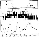 issued from : I.A. McLaren inJ. Fish. Res. Board Can., 1978, 35. [p.1335, Fig.3]. issued from : I.A. McLaren inJ. Fish. Res. Board Can., 1978, 35. [p.1335, Fig.3].
Life cycles of Microcalanus pygmaeus in Loch Striven (55°55'N, 05°10'W).
Relative abundance of C V as a percentage of all copepodids (lower panel); size and numbers per haul (including combined hauls from 60 m to 10 m and 10 to 0 m) of adult females (middle panel), and percentage of nauplii and copepodids above 10 m (from split hauls, taken from 60 to 10 m and 10 to 0 m) (upper panel).
Successive generations as infered from peaks in the C V cohorts and size changes designated as Go, G1, etc.
Data from Marshall (1949, tables V and XII). |
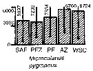 Issued from : A. Atkinson & J.D. Sinclair in Polar Biol., 2000, 23. [p.50, Fig.3] Issued from : A. Atkinson & J.D. Sinclair in Polar Biol., 2000, 23. [p.50, Fig.3]
Microcalanus pygmaeus from Scotia Sea.
Median and interquartile ranges of copepods (nos /m2) in the five water zones; from north to south these are SAF Subantractic Front area, PFZ Polar frontal Zone, PF Polar Front area, AZ Antarctic Zone, WSC Weddell-Scotia Confluence area/ East Wind Drift.
Numbers on the plots are upper interquartiles where these could not be scaled. |
 Issued from : S.B. Schnack-Schiel & E. Mizdalski in Mar. Biol., 1994, 119. [p.362, Fig.4]. Issued from : S.B. Schnack-Schiel & E. Mizdalski in Mar. Biol., 1994, 119. [p.362, Fig.4].
Microcalanus pygmaeus from East Weddell Sea. Cumulative percentage of females in different stages of maturity.
Number below bars: absolute numbers of females inspected.
Late winter/early spring: in 1986; Summer: in 1985; Autumn: in 1992. |
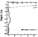 Issued from : P. Ward, A. Atkinson, A.W.A. Murray, A.G. Wood, R. Williams & S. Poulet in Polar Biol., 1995, 15. [p.200, Fig.3]. Issued from : P. Ward, A. Atkinson, A.W.A. Murray, A.G. Wood, R. Williams & S. Poulet in Polar Biol., 1995, 15. [p.200, Fig.3].
Abundance and biomass (g dry mass/m3) profiles for the shelf station (54°48'S, 38°15'W) in January 1990.
Values on the horizontal axes at the base represent abundance and the one above biomass. Solid line = midday haul; hatched line = midnight haul. |
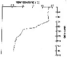 Issued from : P. Ward, A. Atkinson, A.W.A. Murray, A.G. Wood, R. Williams & S. Poulet in Polar Biol., 1995, 15. [p.198, Fig.1, A (modified C.R.)]. Issued from : P. Ward, A. Atkinson, A.W.A. Murray, A.G. Wood, R. Williams & S. Poulet in Polar Biol., 1995, 15. [p.198, Fig.1, A (modified C.R.)].
Temperature profile for the shelf station at Bird Island, South Georgia (54°48'S, 38°15'W) in January 1990. |
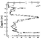 Issued from : P. Ward, A. Atkinson, A.W.A. Murray, A.G. Wood, R. Williams & S. Poulet in Polar Biol., 1995, 15. [p.202, Fig.4, A]. Issued from : P. Ward, A. Atkinson, A.W.A. Murray, A.G. Wood, R. Williams & S. Poulet in Polar Biol., 1995, 15. [p.202, Fig.4, A].
Abundance (10/m3) and biomass (g dry mass/m3) profiles at the oceanic station from the shelf break in water 4000 m deep off Bird Island, South Georgia (53°04'S, 39°51'W) in January 1990.
Values on the horizontal axes at the base of each profile represent abundance and the one above biomass. Solid line = midday haul; hatched line = midnight haul. |
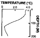 Issued from : P. Ward, A. Atkinson, A.W.A. Murray, A.G. Wood, R. Williams & S. Poulet in Polar Biol., 1995, 15. [p.198, Fig.1, B (modified C.R.)]. Issued from : P. Ward, A. Atkinson, A.W.A. Murray, A.G. Wood, R. Williams & S. Poulet in Polar Biol., 1995, 15. [p.198, Fig.1, B (modified C.R.)].
Profile temperature-depth at the oceanic stations from the shelf break in water 4000 m deep off Bird Island, South Georgia, in January 1990. |
 Issued from : K.M. Swadling, So. Kawaguchi & G.W. Hosie in Deep-Sea Research II, 2010, 57. [p.898, Fig.6 (continued)]. Issued from : K.M. Swadling, So. Kawaguchi & G.W. Hosie in Deep-Sea Research II, 2010, 57. [p.898, Fig.6 (continued)].
Distribution of indicator species Microcalanus pygmaeus from the BROKE-West survey (southwest Indian Ocean) during January-February 2006.
Sampling with a RMT1 net (mesh aperture: 315 µm), oblque tow from the surface to 200 m.
The survey area was located predominantly within the seasonal ice zone, and in the month prior to the survey there was considerable ice coverage over the western section but none over the east.
See map showing sampling sites in Calanus propinquus. |
 Issued from : J. Michels, S.B. Schnack-Schiel, A. Pasternak, E. Mizdalski, E. Isla & D. Gerdes in Polar Biol., 2012, 35. [p.377, Fig.8 (part., copepodid stages omitted.)]. Issued from : J. Michels, S.B. Schnack-Schiel, A. Pasternak, E. Mizdalski, E. Isla & D. Gerdes in Polar Biol., 2012, 35. [p.377, Fig.8 (part., copepodid stages omitted.)].
Vertical distribution of the adult females and males of Microcalanus pygmaeus from East Weddell Sea shelf (70°48.558'S, 10°43.698'W) between 9 and 28 December 2003. The water depth at the sampling spots varied between 438 and 484 m.
The white dots represent the weighted mean depths. |
 Issued from : G.B. Deevey in Bull. Bingham Oceanogr. Coll., 1960, 17 (2). [p.74, Fig.10]. Issued from : G.B. Deevey in Bull. Bingham Oceanogr. Coll., 1960, 17 (2). [p.74, Fig.10].
Obsrrved seasonal variations in median cephalothorax length of female Microcalanus pygmaeus (Marshall & al., 1934) in Loch Striven in 1933) compared with length calculated from the mean temperature and number of diatoms of the month previous to sampling. |
 Issued from : G. B. Deevey in Bull. Bingham Oceanogr. Coll., 1960, 17 (2). [p.73, Table VIII]. Issued from : G. B. Deevey in Bull. Bingham Oceanogr. Coll., 1960, 17 (2). [p.73, Table VIII].
Correlation coefficients for mean cephalothorax lengths with the temperature and phytoplankton of the day of samplingt and averaged for the previous four weeks obtained with 31 and 29 pairs of data for female Microcalanus pygmaeus from Loch Striven (W Clyde estuary)).
Quantity of diatoms (data published by S.M. Marshall, 1949 .
In 1933 the extreme range temperature at 0 m was 4.5-16.4°C and at 30 m 6.37-12.98°C. |
| | | | Loc: | | | Antarct. (Amundsen Sea, Gerlache Strait, Croker Passage, Peninsula, Scotia Sea, Weddell Sea, off Halley Bay, S South Georgia, SW Atlant., Indian, Lützow-Holm Bay, SW & SE Pacif., King George Is., Potter Cove, McMurdo Sound), sub-Antarct. (Prince Edward Is., N South Georgia, SW Atlant., Indian, SE Pacif.), South Africa (E), Namibia, off Mauritania, SW Atlant., off Amazon, Caribbean Sea, Caribbean Colombia, Bahia de Mochima (Venezuela), G. of Mexico, Arct. (Barents Sea, Franz Josef Land, Laptev Sea, Kara Sea, New-Siberia Archipelago, Chukchi Sea, Beaufort Sea, Nansen Basin, Amundsen Basin, Barrow Strait, Makarov Basin, Canadian abyssal plain, Lomonosov Ridge, Bering Sea, St. Lawrence Island, Anadyr Strait, Saguenay fjord, G. of St. Lawrence, off Newfoundland, Cape Flemish, Barrow Strait, Ungava Bay, Ameralik & Godthaab fjords, W Baffin Bay, Greenland Sea, Fram Strait, Kongsfjorden, Faroe Is., Iceland, Spitzbergen, Pechora Sea, Norwegian Sea, Bay of Biscay, North Sea, NW Spain ( La Coruña), Medit. (Ligurian Sea, Tyrrhenian Sea, S Adriatic Sea), Indian ( W, Arabian Sea), Korea, Japan Sea, Japan, off Sanriku, S Kuril Is.,Okhotsk Sea, Station Knot, Bering Sea, S Aleutian Basin, S Aleutian Is., Gulf of Alaska (Icy Strait), British Columbia, off Hawaii, Chile (S, Concepcion, off Santiago), Strait of Magellan | | | | N: | 154 | | | | Lg.: | | | (7) F: 0,9-0,65; M: 1,1; (25) F: 0,78-0,7; (33) F: 0,65-0,78; (35) F: 0,88-0,7; M: 0,8; (36) F: 0,86-0,69; M: 0,84-0,64; (55) F: 0,85; (66) F: 0,60-0,83-0,6; M: 0,76-0,70; (102) F: 0,7; 0,65; M: 0,85; 0,8; (131) F: 0,88-0,7; M: 1,08-0,8; (134) F: ± 0,86; M: ± 1; (196) F: 0,72-0,65; (202) F: 0,7-0,88; M: 0,8; (208) F: 1,12; M: 0,72-0,69; (376) F: 0,77; (432) F: 0,72; (1113) F: 0,58-0,77; (1232) F: 0,6-0,88; M: 0,7-0,8; {F: 0,58-1,12; M: 0,64-1,10} | | | | Rem.: | epi-bathypelagic.
Sampling depth (Antarct., sub-Antarct.) : 0-1000 m.
For Vervoort (1957, p.36) the differences between both forms (M. pygmaeus and M. pusillus originally descrbed by Sars) are mainly localized in the A1 (these reach the genital somite in M. pusillus and the caudal rami in M. pygmaeus and the swimming feet (in M. pusillus the legs are comparatively short and broad, whilst the terminal spines on the 2nd-4th exopods have coarse denticles; in M. pygmaeus the legs are more slender, while the terminal spines of the 2nd-4th exopods have a finely serrated margin). Since Sars' descriptions of both forms intermediate specimens have been recorded from Arctic and boreal localities and its has become clear that the points of difference, as outlined by Sars, are insufficient for specific distinction. It is reasonable to suppose that only a single species: M. pygmaeus. If, for some reason, it is advisable to separate specimens, they can be named M. pygmaeus pusillus (short A1) and M. pygmaeus pygmaeus (long A1).
For Mazzocchi & al. (1995) all intermediate forms collected, seem to demonstrate the uniqueness of this species. Only a genetic analysis could clearly define the extent of the variability. | | | Last update : 17/06/2021 | |
|
|
 Any use of this site for a publication will be mentioned with the following reference : Any use of this site for a publication will be mentioned with the following reference :
Razouls C., Desreumaux N., Kouwenberg J. and de Bovée F., 2005-2026. - Biodiversity of Marine Planktonic Copepods (morphology, geographical distribution and biological data). Sorbonne University, CNRS. Available at http://copepodes.obs-banyuls.fr/en [Accessed February 05, 2026] © copyright 2005-2026 Sorbonne University, CNRS
|
|
 |
 |


