|
|
 |
|
Calanoida ( Order ) |
|
|
|
Diaptomoidea ( Superfamily ) |
|
|
|
Acartiidae ( Family ) |
|
|
|
Acartiella ( Genus ) |
|
|
| |
Acartiella natalensis (Connell & Grindley, 1974) (F,M) | |
| | | | | | | Syn.: | Acartia (Acartiella) natalensis Connell & Grindley, 1974 (p.89, figs.F,M); Wooldridge & Erasmus, 1980 (p.107, tidal utilization); Jerling, 2008 (p.55, Tabl.1); Jerling & al., 2010 (p.543, Table 1, fig.3);
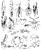
Acartia natalensisKibirige & Perissinotto, 2003 (p.727, Table 1, 2, fig.5, seasonal distribution) |  issued from : A.D. Connell & J.R. Grindley in Ann. S. Afr. Mus., 1974, 65 (3). [p.90, Figs.1-12]. As Acartia (Acartiella) natalensis. Female (from Mtentu River estuary): 1, habitus (dorsal); 3, urosome (dorsal); 6, A2; ; 7 Md (cutting edge); 8, Mxp; ; 9, P1; 11, P5. Nota: A1 22 apparent segments, but 'segment' 2 apparently compounded of 3 segments and 'segment' 4 of 2, giving 25 true segments; segments 13 to 20 (apparently 10 to 17) have a row of small spines Male: 2, habitus (dorsal); 4, urosome (dorsal); 5, geniculate portion of right A1; 10, P4; 12, P5 (posterior); 12 a, c, right P5 (anterior view); 12 b, left P5 (anterior view). Nota: The last urosome segment has a couple of small spines, and there are usually 2 or 3 on the outer edge of the caudal rami; only the left ramus has a short curved accessory seta, whilethe plumose setae are similar tothose of the female.A1 extend to the distal end of the caudal rami; the left A1 is as in the female, while the right has only 17 apparent 'segments', several of them compound; the geniculation is between 'segments 15 and 16 these being compounds of true segments 17-18 and 19-21 respectively; apparent segment 9 bears a short heavy spine, while 10 and 11 are swollen; 'segments' 14, 15 and 16 bear serrated, sabre-like spines. Right P5 does however form a well-developed clasping apparatus (as specified by Sewell, 1914, in his definition of the subgenus); the detail of the terminal structures of left P5 segment 2-3 was difficult to determine clearly (see microphotographs by scanning electon microscope in fig.24-25). Scale bars in microns.
|
 issued from : A.D. Connell & J.R. Grindley in Ann. S. Afr. Mus., 1974, 65 (3). [p.93, Table 1]. As Acartia (Acartiella) natalensis. Ornamentation of the swimming legs P1 to P4. Si, Se, St represent internal, external and terminal spines or setae respectively. Spines in roman numerals; setae in arabic numerals.
|
 issued from : A.D. Connell & J.R. Grindley in Ann. S. Afr. Mus., 1974, 65 (3). [p.94, Figs.24-25]. As Acartia (Acartiella) natalensis. Male: left P5, 24 lateral and 25 dorsal (with some debris caught in the setae).
| | | | | Compl. Ref.: | | | Carrasco & al., 2013 (p.45, turbidity effects vs. feeding) | | | | NZ: | 1 | | |
|
Distribution map of Acartiella natalensis by geographical zones
|
| | | | Loc: | | | Mozambique (estuaries), South Africa (Algoa Bay, Mpenjati Estuary, Mhlathuze Estuary and Richard's Bay Harbour, St Lucia estuary) | | | | N: | 6 | | | | Lg.: | | | (152) F: 1,05-0,82; M: 0,88-0,73; {F: 0,82-1,05; M: 0,73-0,88} | | | Last update : 03/12/2020 | |
|
|
 Any use of this site for a publication will be mentioned with the following reference : Any use of this site for a publication will be mentioned with the following reference :
Razouls C., Desreumaux N., Kouwenberg J. and de Bovée F., 2005-2026. - Biodiversity of Marine Planktonic Copepods (morphology, geographical distribution and biological data). Sorbonne University, CNRS. Available at http://copepodes.obs-banyuls.fr/en [Accessed February 02, 2026] © copyright 2005-2026 Sorbonne University, CNRS
|
|
 |
 |


