|
|
 |
|
Calanoida ( Order ) |
|
|
|
Epacteriscoidea ( Superfamily ) |
|
|
|
Epacteriscidae ( Family ) |
|
|
|
Bunderia ( Genus ) |
|
|
| |
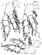
Bunderia misophaga Jaume & Humphreys, 2001 (F,M) | |
| | | | | | | Ref.: | | | Jaume & Humphreys, 2001 (p.158, figs.F,M) | 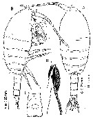 issued from : D. Jaume & W.F. Humphreys in J. Crustacean Biol., 2001, 21 (1). [p.159, Fig.1]. Female (from 22°25'S, 113°46'E): A-B, habitus (dorsal and lateral, respectively); C, genital double-somite (ventral); D, detail of caudal rami (dorsal; showing relative lengths of caudal setae).
|
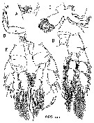
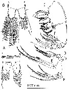 issued from : D. Jaume & W.F. Humphreys in J. Crustacean Biol., 2001, 21 (1). [p.160, Fig.2]. Female: A, detail of anal somite and caudal rami (dorsal); B, idem (ventral; arrow pointing to reduced caudal seta I); C, Mx2 (setae on proximal endopodal segment and ornamentation of setae of distal endopodal segment omitted); D, detail of 1st endopodal segment (pair of setae corresponding to armature of ancestral segment I omitted); E, detail of 2 setae omitted in D; F, detailof 2nd endopodal segment.
|
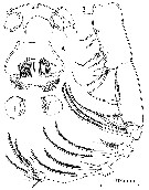 issued from : D. Jaume & W.F. Humphreys in J. Crustacean Biol., 2001, 21 (1). [p.161, Fig.3]. Female: A, rostrum, labrum and paragnaths; B, Mxp.
|
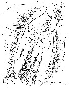 issued from : D. Jaume & W.F. Humphreys in J. Crustacean Biol., 2001, 21 (1). [p.162, Fig.4]. Female: A, A1 (ventral); B, detail of modified seta on 1st segment; C, A2; A2; D, P1 (anterior view); E, detail of posterior surface of inner basal seta of former;
Male: F, right A1 (ventral; arrow pointing to anterodistal spinous process on segment 20; paratype).
|
 issued from : D. Jaume & W.F. Humphreys in J. Crustacean Biol., 2001, 21 (1). [p.165, Fig.5]. Female: A, P2 (anterior); B, P3 (anterior); C, P4 (anterior).
|
 issued from : D. Jaume & W.F. Humphreys in J. Crustacean Biol., 2001, 21 (1). [p.166, Fig.6]. Female: A, Md (cutting edge of coxal gnathobase); B, Md (mandibular palp); C, Mx1; D, left P5 (anterior). Male: P5 (posterior; paratype).
| | | | | NZ: | 1 | | |
|
Distribution map of Bunderia misophaga by geographical zones
|
| | | | | | | Loc: | | | W Australia: Bundera (Cape Range Peninsula: cave) | | | | N: | 1 | | | | Lg.: | | | (815) F: 1,47; 1,37; M: 1,73; {F: 1,37-1,47; M: 1,73} | | | | Rem.: | anchialine. | | | Last update : 30/12/2014 | |
|
|
 Any use of this site for a publication will be mentioned with the following reference : Any use of this site for a publication will be mentioned with the following reference :
Razouls C., Desreumaux N., Kouwenberg J. and de Bovée F., 2005-2026. - Biodiversity of Marine Planktonic Copepods (morphology, geographical distribution and biological data). Sorbonne University, CNRS. Available at http://copepodes.obs-banyuls.fr/en [Accessed June 22, 2026] © copyright 2005-2026 Sorbonne University, CNRS
|
|
|
|


;)
;)
;)
;)
;)
;)

