|
|
 |
|
Calanoida ( Order ) |
|
|
|
Clausocalanoidea ( Superfamily ) |
|
|
|
Euchaetidae ( Family ) |
|
|
|
Paraeuchaeta ( Genus ) |
|
|
| |
Paraeuchaeta investigatoris Sewell, 1929 (F,M) | |
| | | | | | | Syn.: | Paraeuchaeta californica : A. Scott, 1909 (p.71, figs.F); Sewell, 1913 (p.354); 1929 (p.158);
Euchaeta investigatoris : Vervoort, 1957 (p.77);
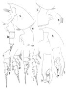
Pareuchaeta investigatoris : Tanaka & Omori, 1968 (p.237, figs.F) | | | | Ref.: | | | Sewell, 1929 (p.158, Descr.M, figs.M); 1947 (p.125, Rem. F,M); Silas, 1972 (p.646); Bradford & al., 1983 (p.23); Park, 1994 (p.320); 1995 (p.59, Rem.F,M, figs.F,M); Chihara & Murano, 1997 (p.800, Pl.106: F) |  issued from : T. Park in Bull. Scripps Inst. Oceanogr. Univ. California, San Diego, 1995, 29. [p.163, Fig.53]. Female: forehead (left side); b, urosome (left); c, d, genital somite (left, right, respectively); e, outer lobe of Mx1; f, P1 (anterior); g, P2 (anterior). Nota: Laterally, rostrum thick, pointing obliquely forward in parallel to anterodorsal margin of forehead; its anterior margin beginning some distance from suprafrontal sensilla and nearly straight. Genital somite with a highly conspicuous dorsal hump about 1/3 its length from proximal end.Genital flange with extraordinarily large posterior lobe, which on left side extending in a posterodorsal direction and which on right side in posteroventral direction. Posterior edge of genital field not produced and merged indistinguishably into posterior margin of genital prominence, which is bulging with more or less irregular outline and a little longer than posterior ventral wall of somite. Cephalosomal appendages similar to those of P. malayensis except that outer lobe of Mx1 with 6 long setae in addition to 2 minute setae proximally. In P1 exopod, outer margin of 1st segment strongly bulging and outer spine very small; outer spine of 2nd segment far short of reaching base of following outer spine. Male: h, forehead (left); i, P1 (anterior); j, P2 (anterior); k, distal exopodal segments of left 5th leg (lateral, tilted clockwise); l, idem (medial, tilted counterclovkwise).
|
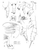
 issued from : O. Tanaka & M. Omori in Publs Seto Mar. Biol. Lab., 1968, XVI (4). [p.238, Fig.11]. As Pareuchaeta investigatoris. Female (from Izu region): A, last thoracic segment and urosome (lateral left side); B, idem (dorsal); C, genital complex (ventral); D, A2; E, Md; F, Mx1; G, Mx2; H, Mxp; I, P1; J, P2. Nota: The urosome segments and furca are in the proportional lengths as 44:21:21:1:13 = 100. The anal segment is concealed beneath the foregoing. Prosome and urosome are in the proportional lengths as 75 to 25. There is slight difference in the number of setae on the outer lobe of the Mx1 with Sewell's (1947) specimen.
|
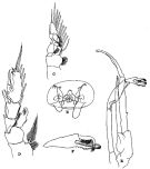
 issued from : R.B.S. Sewell in The John Murray Expedition, 1933-34, Scientific Reports, VIII (1), 1947. [p.124, Fig.28, B-F]. Female (from Arabian Sea): B, genital complex (ventral); C, P1; D, P2. Nota: The proportional lengths of the various segments of the body (cephalon to caudal rami) as 305:126:101:81:68:127:76:68:10:38 = 1000. Head and 1st pediger segment incompletely fused, the chitin splits along the line pf fusion. The postero-lateral region of the fused 4th and 5th thoracic segments is produced baxkwards in a rounded margin, that bears on its inner aspect a somewhat scanty tuft of long hairs. In the A2 the 1st basal segment bears a row of long hairs on its inner margin, and 1 external seta. In Mx1 the number of setae arising from the parts of the appendage: 1st inner lobe (13), 2 nd inner lobe (1), 3 rd inner lobe (1); basal segment 2 (4); endopod 1 (6), endopod 2-3 (11); exopod (11); outer lobe (6 setae). Male: E, P5; F, clasping organ of left P5.
|
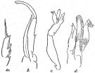
 issued from : R.B.S. Sewell in Mem. Indian Mus., 1929, X. [p.159, Fig.60]. Male (from off SE Sri-Lanka): a, exopdal segment 3 of P2; b, right P5; c, left P5; d, distal portion of left P5 (enlarged).
|
 issued from : A. Scott in Siboga-Expedition, 1909, XIX a. [Plate XV, Figs.1-8]. As Paraeuchaeta californica. Female (from Indonesia-Malaysia): 1, habitus (dorsal); 2, forehead (lateral); 3, last thoracic and genital segments (lefi side); 4, A1; 5, Mxp (part of one of the distal hairs); 6, P1; 7, P2; 8, P3 (part of terminal spine of exopodite). Nota: The figure shewing the lateral view of the genital segment is rather different from that given by Esterly (1906); the protuberances at the side of the genital opening, appear to be more pronounced than in Esterly's type.
|
 Paraeuchaeta investigatoris Paraeuchaeta investigatoris Female: 1 - See key to species Groups and independent species of Paraeuchaeta (p.30): malayensis species Group. 2 - Outer spine of 2nd exopodal segment (or the 2nd of the first 2 exopodal segments forming a proximal, compound segment) of P1 normally developed (Fig.53-f). 3 - Outer lobe of Mx1 with 6 long setae in addition to 1 small and a minute setae proximally (Fig.53-e. 4 - Genital somite without a conical process on left side close to anterior base of genital prominence (Fig.53-c). 5 - Dorsally and ventrally, genital somite symmetrical. 6 - Laterally, genital flange with extremely large posterior lobe (Fig.53-c). 7 - Laterally, genital somite with conspicuous dorsal hump (Fig.53-c).
| | | | | Compl. Ref.: | | | Sewell, 1948 (p.329, 530, 534, 539, 540, 552); Park & Ferrari, 2009 (p.143, Table 8, biogeography) | | | | NZ: | 4 | | |
|
Distribution map of Paraeuchaeta investigatoris by geographical zones
|
| | | | | | | | | | Loc: | | | G. of Aden, Arabian Sea, off S India, off E Sri Lanka, Bay of Bengal, Indonesia-Malaysia, China Seas (South China Sea), Japan (Izu region) | | | | N: | 5 | | | | Lg.: | | | (3) F: 6,6-5,8; M: 6,5-5,4; (5) F: 7; (11) F: 7-6,58; (29) M: 5,62; (63) F: 6,18; {F: 5,80-7,00; M: 5,40-6,50} | | | | Rem.: | Park (1995, p.60) found this species in the South China Sea, Malay Archipelago, and Bay of Bengal. | | | Last update : 27/01/2015 | |
|
|
 Any use of this site for a publication will be mentioned with the following reference : Any use of this site for a publication will be mentioned with the following reference :
Razouls C., Desreumaux N., Kouwenberg J. and de Bovée F., 2005-2026. - Biodiversity of Marine Planktonic Copepods (morphology, geographical distribution and biological data). Sorbonne University, CNRS. Available at http://copepodes.obs-banyuls.fr/en [Accessed February 02, 2026] © copyright 2005-2026 Sorbonne University, CNRS
|
|
 |
 |


