|
|
 |
|
Calanoida ( Order ) |
|
|
|
Arietelloidea ( Superfamily ) |
|
|
|
Heterorhabdidae ( Family ) |
|
|
|
Paraheterorhabdus ( Genus ) |
|
|
|
Paraheterorhabdus ( Sub-Genus ) |
|
|
| |
Paraheterorhabdus (Paraheterorhabdus) longispinus (Davis, 1949) (F,M) | |
| | | | | | | Syn.: | Heterorhabdus longispinus Davis,1949 (p.58, Descr.M, figs.M);
Heterorhabdus (Paraheterorhabdus) robustoides Brodsky, 1950 (1967) (p.346, figs.F,M); ? Tanaka, 1964 a (p.20, Rem.); Vervoort, 1965 (p.118, Rem.); Heptner, 1971 (p.156, 158, figs.F,M, Rem.); Minoda, 1971 (p.38); 1972 (p.326); Morioka, 1972 a (p.314); Gardner & Szabo, 1982 (p.368, 370, figs.F,M);
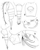
Heterorhabdus robustoides : Hattori, 1991 (tab.1, Appendix); Yamaguchi & al., 2002 (p.1007, tab.1) | | | | Ref.: | | | Park, 2000 (p.76, figs.F,M, Rem.) |  issued from : T. Park in Bull. Scripps Inst. Oceanogr. Univ. California, San Diego, 2000, 31. [p.198, Fig.46]. Female: a, habitus (left side); b, c, urosome (left, dorsal, respectively); d, e, genital somite (ventral, left, respectively); f, masticatory edge of right Md (posterior); g, masticatory edge of left Md (posterior); h, left Mx2 (posterior).
|
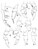
 issued from : T. Park in Bull. Scripps Inst. Oceanogr. Univ. California, San Diego, 2000, 31. [p.199, Fig.47]. Female: a, right Mxp (anterior); b, exopod of P5 (anterior). Male: c, P5 (anterior); d, P5 (endopods omitted), posterior; e, exopod of right P5 (anterior); f, idem (posterior, tilted clockwise); g, h, exopod of left P5 (posterior, anterior).
|
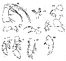
 issued from : C.C. Davis in Univ. Wash. Publs Biol., 1949, n. ser. 14. [Pl.10, Figs.126-127]. As Heterorhabdus longispinus. Male (from NE Pacific): 126, Mxp; 127, left P5 (anterior view).
|
 issued from : C.C. Davis in Univ. Wash. Publs Biol., 1949, n. ser. 14. [Pl.11, Figs.128-137]. As Heterorhabdus longispinus. Male: 128, right P5 (posterior face); 129, endopod of right P5; 130, exopod of right P5 (lateral view from right side); 131, Mx2; 132, right Md (mandibular palp); 133, right Md (masticatory blade); 134, left Md (masticatory blade); 135, P2; 136, P3; 137, exopod of P1. Nota : Head and 1 st thoracic segment separated. The head constitutes more than half the length of the metasome. Front with a small papilla, on the ventral side of which is the rostrum. Lateral angles of the end of prosome are rounded. Urosome slightly greater than ½ the length of the metasome, and all of the five segments more or less equal in length except the anal, which is only 1/3 to ¼ the length of the others. Left caudal ramus longer than the right, and the 2 nd from the inner seta heavier than the others and longer than the entire body of the animal. A1 reach just about to the end of the caudal rami ; right A1 25-segmented ; left one geniculate of 21 segments. 1 st endopodal segment of A2 with a dense row of fine hairs on the distal portion of the outer margin. The masticatory portion of left M dis not the same as that of the right. Mxp with a short thin seta on the middle of the inner border of the 1 st basal segment ; the proximal end of the outer border of the 2 nd segment bears a swelling that is larger than ordinary. P1 with a long spine on the outer distal corner of the 1 st exopodal segment. Protrusion on the inner border of the 2 nd exopodal segment of the right P5 is pointed, and relatively large ; the 2 nd segment is greatly enlarged on the inner border ; the spine on the outer distal corner of the 1st segment of the 1st exopod is very long.
|
 Paraheterorhabdus (Paraheterorhabdus) longispinus Paraheterorhabdus (Paraheterorhabdus) longispinus female: 1 - Left caudal ramus distinctly longer than right and completely fused with anal segment (Fig.46-c) 2 - Dorsally, posterolateral corners of prosome angular (Fig.46-c). 3 - Laterally, 2nd urosomal somite (Fig.46-b) without a swelling along ventral margin. 4- Laterally, posterior slope of genital prominence concave (Fig.46-e).
|
 Paraheterorhabdus (Paraheterorhabdus) longispinus Paraheterorhabdus (Paraheterorhabdus) longispinus female: 1 - Left caudal ramus distinctly longer than right and completely fused with anal segment. 2 - Basis of right P5 with conical inner lobe. 3 - P5 with relatively slender exopods; medial projection of 2nd exopodal segment of right P5 without a lobe on distal side. 4 - Medial projection of 2nd exopodal segment of right P5 toothlike (Fig.47-e, f). 5 - In 3rd exopodal segment of left P5 (Fig.47-d), inner spine about on the same level as outer spine.
| | | | | Compl. Ref.: | | | Galbraith, 2009 (pers. comm.); Park & Ferrari, 2009 (p.143, Table 8, fig.2, biogeography) | | | | NZ: | 4 | | |
|
Distribution map of Paraheterorhabdus (Paraheterorhabdus) longispinus by geographical zones
|
| | | | Loc: | | | Japan, off Sanriku, Station Knot, Kuril-Kamtchatka Trench, Bering Sea, G. of Alaska, NE Pacif. (off Cape Flattery), California | | | | N: | 7 | | | | Lg.: | | | (22) F: 5-4,8; M: 4,8-4,6; (208) F: 5; M: 5,3-4,4; (259) M: 4,3; (824) F: 5,33-4,5; M: 5,08-4,64; {4,50-5,33; M: 4,30-5,08} | | | | Rem.: | Between 0-1100 m. | | | Last update : 27/01/2015 | |
|
|
 Any use of this site for a publication will be mentioned with the following reference : Any use of this site for a publication will be mentioned with the following reference :
Razouls C., Desreumaux N., Kouwenberg J. and de Bovée F., 2005-2026. - Biodiversity of Marine Planktonic Copepods (morphology, geographical distribution and biological data). Sorbonne University, CNRS. Available at http://copepodes.obs-banyuls.fr/en [Accessed February 20, 2026] © copyright 2005-2026 Sorbonne University, CNRS
|
|
 |
 |


