|
|
 |
|
Calanoida ( Order ) |
|
|
|
Hyperbionychidae ( Family ) |
|
|
|
Hyperbionyx ( Genus ) |
|
|
| |
Hyperbionyx pluto Ohtsuka, Roe & Boxshall, 1993 (F,M) | |
| | | | | | | Ref.: | | | Ohtsuka & al., 1993 a (p.70, figs.F,M); Boxshall & Halsey, 2004 (p.129: figs.F,M) | 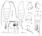 issued from : S. Ohtsuka, H.S.J. Roe & G.A. Boxshall inSarsia, 1993, 78. [p.71, Fig.1]. Female (from off N Cape Verde Islands): A, habitus (dorsal); B, idem (lateral right side); C, genital double somite (ventral); D, rostrum; E, labrum and paragnath (lateral view); F, idem (ventral view); G, paragnath; H, anal somite and caudal rami (ventral). Scales in mm. Paratype: A, B, E, F; holotype: C, D, G, H. Nota: Rostrum triangular, with pair of long filaments. A1 asymmetrical, left longer than right (terminal segments of left A1 missing); right A1 27-segmented. Genital apertures asymmetrically located (left aperture more anteriorly located than right). Caudal rami asymmetrical, right longer than left.
|
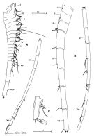 issued from : S. Ohtsuka, H.S.J. Roe & G.A. Boxshall inSarsia, 1993, 78. [p.72, Fig.2]. Female: A, right A1; B, left A1. Male: C, terminal fused segment of left A1. Scales in mm. Holotype: A,B; paratype: C.
|
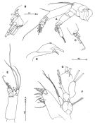 issued from : S. Ohtsuka, H.S.J. Roe & G.A. Boxshall inSarsia, 1993, 78. [p.73, Fig.3]. Female: A, A2; B, terminal part of 2nd endopod segment of A2 (vestigial seta indicated by arrowhead); C, 9th exopod segment of A2; D, Md (mandibular cutting edge); E, Md (mandibular palp); F, Mx1; G, terminal part of exopod of Mx1. Nota: Mandibular gnathobase heavily chitinized, with 2 stout teeth and row of setules. Mx1 with praecoxal arthrite produced medially, bearing 7 spines and 6 setae; coxal endite developed with 3 setae terminally; coxal epipodite with 2 long, plumose setae; 1st basal endite rudimentary; endopod elongate, 1-segmented, with 1 middle, 1 subterminal and 3 terminal setae; exopod expanded, incompletely fused to basis, with 2 plumose setae terminally plus 1 tube pore (at base of outer seta). Scales in mm.
|
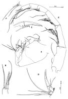 issued from : S. Ohtsuka, H.S.J. Roe & G.A. Boxshall inSarsia, 1993, 78. [p.75, Fig.4]. Female: A, Mx2; B, 2nd coxal endite of Mx2; C, basal endite of Mx2; D, endopod of Mx2; E, Mxp (with the locations of pores indicated by arrowheads); F, 5th endopod segment of Mxp. Nota: Mx2 well developed, 1st praecoxal endite with 3 setae and 2 small, spiniform elements, 2nd with 1 serrate; 1st coxal endite with 2 serrate setae, 2nd coxal endite (Fig.4B) produced anteriorly, with 1 strongly serrate spine and 2 setae terminally; basal endite (Fig.4C) considerably produced anteriorly, with 1 large serrate spine terminally and 1 moderate serrate seta, 2 weak setae and 1 tube pore subterminally; endopod (Fig.4D) indistinctly 4-segmented, 1st well developed, with 1 heavily chitinized spine and 3 weak setae, 2nd with 3 setae, 3rd with 1 seta, 4th with 2 setae. Scales in mm.
|
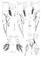 issued from : S. Ohtsuka, H.S.J. Roe & G.A. Boxshall inSarsia, 1993, 78. [p.76, Fig.5]. Female: A, P1 (anterior); B, distal two endopod segments of left P1; C, outer margin of 2nd and 3rd exopod segments of P1; D, P2 (anterior); E, P3 (anterior); F, basal seta of P3; G, P4 (anterior); H, P5 (anterior). Scales in mm.
|
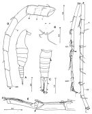 issued from : S. Ohtsuka, H.S.J. Roe & G.A. Boxshall inSarsia, 1993, 78. [p.77, Fig.6]. Male: A, habitus (lateral right side); B, rostrum; C, urosome (dorsal, gonopore indicated by large arrowhead, position of missing caudal seta by small arrowhead); D, left A1 (geniculation indicated by arrowhead); E, ancestral segments XIX to XXI (geniculation indicated by arrowhead). Scales in mm. Nota: Rostrum as in female. Urosome 5-segmented. Genital somite with gonopore. Left A1 geniculate, indistinctly 26-segmented, geniculation between 20th and 21st segments. ventrolaterally on right side.
|
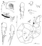 issued from : S. Ohtsuka, H.S.J. Roe & G.A. Boxshall in Sarsia, 1993, 78. [p.79, Fig.7]. Male: A, terminal end of syncoxa of left Mxp; B, terminal end of basis of right Mxp; C, 3rd exopod segment of left P2; D, endopod of left P3; E, 3rd exopod segment of right P3; F, endopod of right P4; G, 3rd exopod segment of left P4; H, P5 (anterior); I, vestigial endopod of left P5; J, terminal seta of 3rd exopod segment of left P5; K, medial spine on 2nd exopod segment of left P5; L, vestigial endopod of right P5. Scales in mm.
|
 issued from : S. Ohtsuka, H.S.J. Roe & G.A. Boxshall inSarsia, 1993, 78. [p.79, Fig.7]. Female (SEM micrographs): A, genital double somite (ventral); B, right genital aperture and folds marking plane of fusion between genital and 1st abdominal somites indicated by arrow. Scale bars: A: 0.2 mm, B: 0.1 mm.
| | | | | Compl. Ref.: | | | Bradford-Grieve, 2004 (p.283) | | | | NZ: | 1 | | |
|
Distribution map of Hyperbionyx pluto by geographical zones
|
| | | | Loc: | | | NE Atlant. (Cape Verde Is.) | | | | N: | 1 | | | | Lg.: | | | (400) F: 10,84-9,96; M: 8,4; {F: 9,96-10,84; M: 8,40} | | | | Rem.: | hyperbenthic at depth (? 4000 m).
See remarks in H. athesphatos | | | Last update : 06/01/2015 | |
|
|
 Any use of this site for a publication will be mentioned with the following reference : Any use of this site for a publication will be mentioned with the following reference :
Razouls C., Desreumaux N., Kouwenberg J. and de Bovée F., 2005-2026. - Biodiversity of Marine Planktonic Copepods (morphology, geographical distribution and biological data). Sorbonne University, CNRS. Available at http://copepodes.obs-banyuls.fr/en [Accessed January 06, 2026] © copyright 2005-2026 Sorbonne University, CNRS
|
|
 |
 |


