|
|
 |
|
Calanoida ( Order ) |
|
|
|
Lucicutiidae ( Family ) |
|
|
|
Lucicutia ( Genus ) |
|
|
| |
Lucicutia maxima Steuer, 1904 (F,M) | |
| | | | | | | Syn.: | Lucicutia grandis : Grice & Hülsemann, 1965 (p.224); no L. maxima : A. Scott, 1909 (p.127, figs.M); Wolfenden, 1911 (p.318); Sars, 1925 (p.211, figs. F); Sewell, 1932 (p.295, figs.M); Rose, 1933 a (part., p.193, Rem.F, figs. F); ? Gunther & Hardy, 1935 (1936) (p.179, Rem.); Tanaka, 1963 (p.49, figs.M); De Decker & Mombeck, 1964 (p.13); Tanaka & Omori, 1967 (p.240) | | | | Ref.: | | | Steuer, 1904 (p.596, Descr.F, fig.F, non M); Wolfenden, 1905 a (p.22, Rem.); ? Farran, 1908 b (p.61, Rem.); Sewell, 1932 (p.295, figs.F, no M); Rose, 1933 a (p.194, Rem.M, fig.M); Hülsemann, 1966 (p.729, figs.F,M, Rem.); Bradford-Grieve & al., 1999 (p.883, 945, figs.F,M); Boxshall & Halsey, 2004 (p.132: F; p.133: M; p.131: figs.F,M); Vives & Shmeleva, 2007 (p.342, figs.F,M, Rem.) | 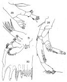 issued from : K. Hülsemann in Bull. Mar. Sc., 1966, 16 (4). [p.710, Figs.28-31]. Female: 28, A2; 29, Md (mandibular palp); 30, Md (biting edge); 31, Mx1.
|
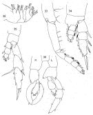 issued from : K. Hülsemann in Bull. Mar. Sc., 1966, 16 (4). [p.712, Figs.32-36]. Female: 32, Mx2; 33, Mxp; 34, P1; 35, P5. Male: 36, P5.
|
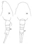 issued from : K. Hülsemann in Bull. Mar. Sc., 1966, 16 (4). [p.716, Figs.55-58]. Female: head (dorsal); 56, idem (lateral); 57, urosome (dorsal); 58, idem (lateral right side). Nota: Spine-like protrusions on anterior of head smaller, directed downward. All females examined had a large anal flap (not shown by Steuer), anal segment swollen. Furca shorter than abdomen.
|
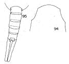 issued from : K. Hülsemann in Bull. Mar. Sc., 1966, 16 (4). [p.722, Figs.94-95]. Male: 94, head (dorsal); 95, urosome (dorsal). Nota: Head with two small pointed protrusions on each side. Abdomen and furca one and one-half times contained in cephalothorax.. length of furca 2/3 of abdomen; furcal rami about 7 times longer than wide. Anal segment slightly shorter than preceding segment. Right A1 exceeding end of furca by 2 segments. 2nd basipodal segment of P1 with tube-like protrusion; endopod of P1 3-segmented. 2nd basipodal segment of right P5 straight with minute teeth on inner margin; 2 nd basipodal segment of left P5 protruded on distal inner corner with small teeth.
|
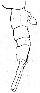 issued from : A. Steuer in Zool. Anz., 1904, 27 (19. [p.596, Fig.4]. Female: last thoracic segment and urosome (lateral).
|
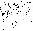 issued from : R. Stephen, 2007 : Data sheets of NIO, Kochi, India (on line). Female (from Indian Ocean): a, habitus (dorsal); b, P1; d, rostrum. Male: c, P5.
|
 Lucicutia maxima Lucicutia maxima female: 1 - Characters following not combined : Prosome about 3 times longer than urosome. Cephalosome with slightly projecting and rounded anterior corners and well developed lateral spinous projections; anal somite about as long as wide; caudal rami 11.7 times longer than wide and bowed outwards at base, leaving elliptical space between rami proximally. 2 - P1 with 3-segmented endopod. 3 - Cephalosome with 2 pairs of spinous projections (1 anterolateral and 1 lateral). 4 - Anterolateral projections small, directed ventrally. Lateral margins of anal somite convex. Caudal rami shorter than urosome.
|
 Lucicutia maxima Lucicutia maxima male: 1 - P1 with 3-segmented endopod. 2 - Cephalosome with 2 pairs of spinous projections (1 anterolateral and 1 lateral). 3 - Anterolateral projections small; lateral projections directed ventrally. 4 - Anal somite slightly shorter than preceding somite; caudal rami about 7 times longer than wide.
| | | | | Compl. Ref.: | | | Grice, 1963 a (p.496); Grice & Hulsemann, 1967 (p.18); 1968 (tab.2); Vinogradov, 1968 (1970) (p.277); Björnberg, 1973 (p.344, 387); Frontier, 1977 a (p.15); Madhupratap & Haridas, 1990 (p.310, fig.3, vertical distribution night/day; fig.7: cluster); Shih & Young, 1995 (p.70); Suarez-Morales & Gasca, 1998 a (p.110); Fernandes, 2008 (p.465, Tabl.2); Salah S. & al., 2012 (p.155, Tableau 1); El Arraj & al., 2017 (p.272, table 2, seasonal composition); | | | | NZ: | 7 + 1 doubtful | | |
|
Distribution map of Lucicutia maxima by geographical zones
|
| | | | | | | | | | | |  Issued from : M. Madhupratap & P. Haridas in J. Plankton Res., 12 (2). [p.310, Fig.3]. Issued from : M. Madhupratap & P. Haridas in J. Plankton Res., 12 (2). [p.310, Fig.3].
Vertical distribution of calanoid copepod (mean +1 SE), abundance No/100 m3. 12- Lucicutia maxima.
Night: shaded, day: unshaded.
Samples collected from 6 stations located off Cochin (India), SE Arabian Sea, November 1983, with a Multiple Closing Plankton Net (mesh aperture 300 µm), in vertical hauls at 4 depth intervalls (0-200, 200-400, 400-600, 600-1000 m). |
| | | | Loc: | | | ? Antarct., sub-Antarct., off E Canary Is., off Morovccan Atlantic Coast, Cap Ghir (Morocco), G. of Mexico, Bermuda, ? Lisbon, ? Ireland, Madagascar (Nosy Bé), W Indian, Bay of Bengal, Andaman Sea, China Seas (South China Sea), SE Pacif., off Juan Fernandez Is., N-S Chile (also sub-Antarct.) | | | | N: | 20 | | | | Lg.: | | | (21) F: 9,3-7,8; M: 7,8-7,7; (1034) F: 8,7; (1068) M: 7,4; {F: 7,80-9,30; M: 7,70-7,80}
| | | | Rem.: | Bathypelagic.
For Hülsemann (1966, p.729) the species described under the name L. maxima by Sars (1925) differs from Steuer's species and also from L. aurita in the relative length of the caudal rami, which are nearly as long as the abdomen,
and in the elongate anal segment. For this reason L. sarsi is proposed for the females described by Sars as L. maxima.
Sewell (1932, fig.94) figures a male which because of its obvious similarity to Sars' (1925, 1924: Pl57, fig.1) female must belong to the same species (= L. sarsi). Sewell's description does not agree with his figure (in the figure the furca is as long as the abdomen) which agrees with sarsi, and the description with aurita. For Hülsemann, Sewell found males of two species: the one shown in figure 94 (= sarsi, the other one described (p.295-297) (= aurita).
Because of the synonymies certain geographic distributions are to be confirmed. | | | Last update : 24/10/2022 | |
|
|
 Any use of this site for a publication will be mentioned with the following reference : Any use of this site for a publication will be mentioned with the following reference :
Razouls C., Desreumaux N., Kouwenberg J. and de Bovée F., 2005-2026. - Biodiversity of Marine Planktonic Copepods (morphology, geographical distribution and biological data). Sorbonne University, CNRS. Available at http://copepodes.obs-banyuls.fr/en [Accessed February 27, 2026] © copyright 2005-2026 Sorbonne University, CNRS
|
|
 |
 |


