|
|
 |
|
Calanoida ( Order ) |
|
|
|
Lucicutiidae ( Family ) |
|
|
|
Lucicutia ( Genus ) |
|
|
| |
Lucicutia parva Grice & Hulsemann, 1965 (F,M) | |
| | | | | | | Syn.: | Lucicutia ovalis : Grice & Hulsemann, 1965 (part.., p.224) | | | | Ref.: | | | Grice & Hülsemann, 1965 (p.224, 244, figs.F,M); Hülsemann, 1966 (p.733, figs.F,M, Rem.); Boxshall & Halsey, 2004 (p.132: F; p.134: M); Vives & Shmeleva, 2007 (p.355, figs.F,M, Rem.) | 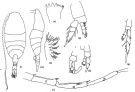 issued from : G.D. Grice & K. Hulsemann in J. Zool., 1965, 146. [p.243, Fig.16, l-s]. Female (from Madeire-Canary Is.): l, habitus (dorsal); m, idem (lateral left side); n, Md (cutting edge); o, Mx2: p, P1; q, P5; Nota: Head and 1st thoracic segment separate, 4th and 5th fused. Rostral filaments slender and short. cephalothorax : urosome ratio about 3 : 1. A1 (25-segmented) exceeds end of furca by at least 4 segments. 2nd basal segment of P1 with tubular protuberance. Anal segment longer than preceding. Furca about 3-times longer than wide Male: left A1 (geniculate portion); s, P5. Nota: Left A1 geniculate (21-segmented), segment 18 with small lamella, 17 with long lamella. Exopod of left P5 with 1 small external spine on 1st segment, 1 short seta on 2nd; 5 setae on 3rd segment of endopod. Right P5 endopod consists of 2 segments of about equal size, the 2nd bears 5 setae.
|
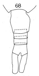 Issued from : K. Hülsemann in Bull. Mar. Sc., 1966, 16 (4). [p.718, Fig.68]. Female: 68, urosome (dorsal).
|
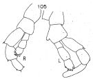 Issued from : K. Hülsemann in Bull. Mar. Sc., 1966, 16 (4). [p.724, Fig.105]. Male: 105, P5. Nota: Inner margin of 2nd basal segment of the right P5 smooth; left P5 with a protrusion about in its middle part bearing minute teeth.
|
 Lucicutia parva Lucicutia parva female: 1 - Characters following not combined : Prosome about 3 times longer than urosome. Cephalosome with slightly projecting and rounded anterior corners and well developed lateral spinous projections; anal somite about as long as wide; caudal rami 11.7 times longer than wide and bowed outwards at base, leaving elliptical space between rami proximally. 2 - P1 with 3-segmented endopod. 3 - Cephalosome without spinous projections. 4 - Genital double-somite symmetrical (dorsal view). 5 - Anal somite much shorter than caudal ramus. 6 - P5 with 3-segmented endopod. 7 - Distal 3 to 4 segments of A1 reaching beyond tip of caudal ramus, body length less than 3 mm.. 8 - Prosome more than twice as long as urosome. 9 - Caudal rami 3 to 4 times longer than wide.
|
 Lucicutia parva Lucicutia parva male: 1 - P1 with 3-segmented endopod. 2 - Cephalosome without lateral projections. 3 - Right A1 reaching 3 to 4 segments beyond tip of caudal rami. 4 - Caudal rami about 4 times longer than wide; inner margin of basis of left P5 protruded in middle, ornamented with several minute teeth; exopod of right P5 3-segmented.
| | | | | Compl. Ref.: | | | Grice & Hulsemann, 1967 (p.18); Park, 1970 (p.477); Medellin-Mora & Navas S., 2010 (p.265, Tab. 2) | | | | NZ: | 4 | | |
|
Distribution map of Lucicutia parva by geographical zones
|
| | | | | | | Loc: | | | off Mauritania, E Azores, W Madeira - Canary Is., Caribbean Sea, Caribbean Colombia, G. of Mexico, Indian, E Pacif. | | | | N: | 5 | | | | Lg.: | | | (21) F: 1,3-1,1; M: 1,2-1; (226) F: 1,29-1,17; M: 1,05; {F: 1,10-1,30; M: 1,00-1,20}
| | | | Rem.: | bathypelagic-hadopelagic.
For Grice & Hulsemann (1965, p.245) this species is related to L. flavicornis. The female differs from the latter in its anal segment which is longer than the preceding one, the A1 is longer than the body (it reaches the furca in flavicornis, see also the length of the terminal seta of the P5 exopod. The male differs from other species in the segmentation of the right P5 (exopod with 3 segments in contrast to 2 segments in other species of the genus). | | | Last update : 16/01/2015 | |
|
|
 Any use of this site for a publication will be mentioned with the following reference : Any use of this site for a publication will be mentioned with the following reference :
Razouls C., Desreumaux N., Kouwenberg J. and de Bovée F., 2005-2024. - Biodiversity of Marine Planktonic Copepods (morphology, geographical distribution and biological data). Sorbonne University, CNRS. Available at http://copepodes.obs-banyuls.fr/en [Accessed November 21, 2024] © copyright 2005-2024 Sorbonne University, CNRS
|
|
|
|


;)
;)
;)

