|
|
 |
|
Calanoida ( Order ) |
|
|
|
Metridinidae ( Family ) |
|
|
|
Metridia ( Genus ) |
|
|
| |
Metridia ferrarii Markhaseva, 2001 (F,M) | |
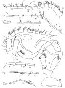
| | | | | | | Ref.: | | | Markhaseva, 2001 (p.44, figs.F,M); Andronov, 2014 (p.45, fig.: P1; Rem.: p.46) | 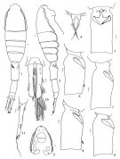 issued from : E.L. Markhaseva in Zoosyst. Rossica, 2001, 9 (1). [p.51, Figs.1-11]. Female (from 69°06'S, 95°02'W): 1, 2, habitus (dorsal and left lateral); 3, cephalosome (ventral view); 4, rostrum; 5, genital somite (ventral view); 6-9, the same (right lateral view); 10, 11, caudal rami (dorsal and right lateral view), from different stations. Nota : Prosome 1.06-1/25 times as long as urosome. Cephalosome with collar-like extension of both left and right lateral margin. Rostrum of two filaments withsetules at the subdivided base. Genital somite nearly twice as long as wide, with elongate spermathecae, left one often darker. Caudal rami about 6-7 times as long as wide.
|
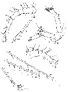
 issued from : E.L. Markhaseva in Zoosyst. Rossica, 2001, 9 (1). [p.54, Figs.27-31]. Female: 27, Md (gnathobase); 28, first leg; 29, fifth legs; 30, 31, cephalosome (lateral and dorsal views). P1 basipod with curved anterior setae and 1 lateral distal seta ; endopod segment 1 with row of spinules in the medial distal corner ; endopod segment 2 with semicircular sclerotized ridge. Endopod segment 1 of P2 with 2 well developed hook-like spines, distal one subdivided, with horns nearly equal in length.
|
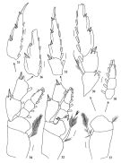 issued from : E.L. Markhaseva in Zoosyst. Rossica, 2001, 9 (1). [p.55, Figs.32-39]. Female: 32, second leg (exopod 3 omitted); 33, exopod 2-3 of second leg 34, third foot (exopod 3 omitted); 35-36, exopod 3 of third leg; 37, coxopod and basipod of fourth leg; 38, endopod of fourth leg; 39, exopod of fourth leg. P1 basipod with curved anterior setae and 1 lateral distal seta ; endopod segment 1 with row of spinules in the medial distal corner ; endopod segment 2 with semicircular sclerotized ridge. Endopod segment 1 of P2 with 2 well developed hook-like spines, distal one subdivided, with horns nearly equal in length.
|
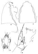
 issued from : E.L. Markhaseva in Zoosyst. Rossica, 2001, 9 (1). [p.56, Figs.40-46]. Male: 40, 41, habitus (dorsal and left lateral); 42, cephalosome (dorsal view); 43, cephalosome (right lateral view); 44, cephalosome (ventral view); 45, posterior prosomal and genital somite (dorsal view); 46, caudal rami (dorsal view). Nota : Prosome 1.08-1.20 times as long as urosome. Lateral collar not developed. Genital somite with small projection on the left covered with spinules. Caudal rami about 7 times as long as wide. Left A1 24-segmented, reaching the end of caudal rami (or exceeding it by 2 distal segments) ; right A1 21-segmented, geniculated, reaching the middle length or the end of caudal rami. Oral parts and swimming legs as in females.
|
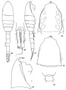
 issued from : E.L. Markhaseva in Zoosyst. Rossica, 2001, 9 (1). [p.57, Figs.47-59]. Male: 47, left A1 (segments 1-6; 48, left A1 (segments 7-11); 49, left A1 (segments 12-15); 50, left A1 (segments 16-19); 51, left A1 (segments 20-24); 52, right A1 (segments 1-12); 53, right A1 (segments 13-15); 54, right A1 (segments16-18); 55, right A1 (segments 19-21); 56, fifth legs; 57, exopod 3 of right foot; 58, right fifth leg (coxopod, basipod and exopod 1-2); 59, left fifth leg (exopod 2-3). Nota : P5 with spinules at coxopod on the left longer segment ; left basipod with spinules in the medial distal part . Left and right basipods with lateral setae supplied with setules distally. Left P5 exopod segment 1 with small spine in lateral distal part of the segment ; exopod segment 2 with hairs proximally and 1 proximal setule ; exopod segment 3 with 2 distal setules ; right P5 seopod segment 1 with small lateral spine and long attenuation exceeding the length of exopod segment 2, the latter supplied with tiny spinule in proximal half of the segment ; exopod segment 3 with 1 and 2 tiny spinules in distal part.
|
 issued from : E.L. Markhaseva in Zoosyst. Rossica, 2001, 9 (1). [p.52, Figs.12-18]. Female: 12, left A1 segments 1-8); 13, idem (segments 9-12); 14, idem (segments 13-15); 15, idem (segments 13-15); 15, idem (segments 21-24); 17, right A1 (segments 1-6); 18, A2. Nota : A1 24-segmented, exceeding the body length by 3-5 distal segments, setal elements are setae and aesthetascs, often difficult to distinguish between them as many setae are weakly sclerotized and apparently transformed into aesthetascs.
|
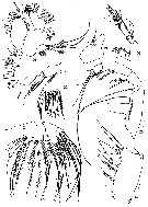 issued from : E.L. Markhaseva in Zoosyst. Rossica, 2001, 9 (1). [p.53, Figs.19-26]. Female: 19, Md (basis); 20, Md (mandibular palp: exopodite and endopodite); 21, iiner lobe 1, inner lobe 4, endopod, exopod and outer lobe 2; 22, Mx1 (inner lobe 1 and inner lobe 2); 23, Mx1 (outer lobe 2); 24, Mx2; 25, Mxp (syncoxa); 26, Mxp (basis and endopod. Scale bars: 0.1 mm. Nota : Md basis with 4 setae ; endopod segments 1 and 2 with 4 and 8 terminal setae plus 2 posterior setae, respectively. Mx1 : praecoxal arthrite (= 1st lobe ) with 9 terminal, 4 posterior and 2 anterior setae ; coxal endite (= 2nd lobe with 5 setae ; basal endites ( = lobes3 and 4) with 4 and 5 setae resoectively ; endopod with 6+11 setae ; exopod with 11 setae ; basal exite ( = 2nd external lobe) with 7+2 setae. Mx2 : with 4+5 setae at inner lobe 1 (praecoxal endite), inner lobe 2 and inner lobes 3-4 (= coxal endites) with 3 setae eachinner lobes 5-6 (basal endites) with 4 setae each ; exopod with 7 setae. Mxp syncoxa proximal to distal with 1, 2, 4 and 4 setae in distal froup and lateral distal seta ; basis with 3 medial setae and row of spinules at their base ; 2 setae distally at endopod segment 1 which is incompletely incorporated into basis ; endopod 5-segmented with 4, 4, 3, 3 and 4 setae
|
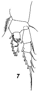 Issued from : V.N. Andronov in Russian Acad. Sci. P.P. Shirshov Inst. Oceanol. Atlantic Branch, Kaliningrad, 2014. [p.45, Fig.13, 7]. Metridia ferrarii after Markhaseva, 2001.
| | | | | Compl. Ref.: | | | Park & Ferrari, 2009 (p.143, Table 4, Appendix 1, biogeography) | | | | NZ: | 3 | | |
|
Distribution map of Metridia ferrarii by geographical zones
|
| | | | | | | | | | Loc: | | | Antarct. (Atlant., Pacif., Indian), Indian (off N Rodriguez Is.) | | | | N: | 1 | | | | Lg.: | | | (823) F: 9,7-8,8; M: 3,8-3,31; {F: 8,80-9,70; M: 8,75-8,95} | | | | Rem.: | Bathy-abyssopelagic. | | | Last update : 20/04/2016 | |
|
|
 Any use of this site for a publication will be mentioned with the following reference : Any use of this site for a publication will be mentioned with the following reference :
Razouls C., Desreumaux N., Kouwenberg J. and de Bovée F., 2005-2026. - Biodiversity of Marine Planktonic Copepods (morphology, geographical distribution and biological data). Sorbonne University, CNRS. Available at http://copepodes.obs-banyuls.fr/en [Accessed June 14, 2026] © copyright 2005-2026 Sorbonne University, CNRS
|
|
|
|


;)
;)
;)
;)
;)
;)
;)
;)

