|
|
 |
|
Calanoida ( Order ) |
|
|
|
Clausocalanoidea ( Superfamily ) |
|
|
|
Aetideidae ( Family ) |
|
|
|
Azygokeras ( Genus ) |
|
|
| |
Azygokeras columbiae Koeller & Littlepage, 1976 (F,M) | |
| | | | | | | Ref.: | | | Koeller & Littlepage, 1976 (p.1548, figs.F,M); Gardner & Szabo, 1982 (p.256, figs.F,M); Markhaseva, 1996 (p.47, figs.F,M) | 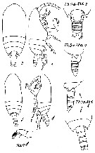 Issued from : E.L. Markhaseva in Proc. Zool. Inst., RAN, 1996; [p.48, Fig.30]. After Koeller & Littlepage, 1976. Female and Male, habitus and urosome.
|
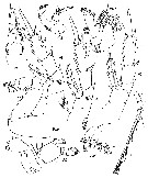 Issued from : E.L. Markhaseva in Proc. Zool. Inst., RAN, 1996. [p.49, Fig.31]. After Koeller & Littlepage, 1976. Female.
|
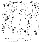 Issued from : P.A. Koeller & J.L. Littlepage in J. Fish. Red. Board Can., 1976, 33. [p.1549, Fig.1]. Female (from 50°27'N, 125°04.5'W): A, posterior part of body (lateral); B, A1; C, Md; D, habitus (lateral); E, Mx1; F, Mxp; G, habitus (dorsal); H, Mx2; I, posterior part of body (dorsal); J, part of terminal spine on exopod of swimming leg; K, A2. Nota: - Maetasome about 3.5 times length of urosome. - Rostrum absent, but 2 minute sensory hairs present on forehead. - Last thoracic segment produced posteriorly into acute points reaching to about midlength of genital segment; points directed posteriorly. - Genital segment large, almost spherical. - 2nd and 3rd abdominal segments about equal in length. - Anal segment shortest. Caudal rami slightly longer than wide, with 4 long terminal setae, 1 short external seta, and 1 short ventral seta. - A1 24-segmented, reaching about to the anal segment. - Basipod of A2 with 4 setae, 2 placed proximally, 2 distally. Exopod 7-segmented. Endopod 2-segmented; 2nd segment with 14 setae in groups of 3, 5, and 6. Md with strong masticatory blade (gnathobase). 2nd basipod with 3 internal setae; 1st endopodal segment with 2 setae; distal end of 2nd endopodal segment with 10 setae. - Mx1 with 1st inner lobe having 7 stout spines, 2 less stout, more plumose proximal spines, and 1 large and 1 small seta. Some minute spinules present on 1st inner lobe. 2nd and 3rd inner lobes with 4 setae each, 3rd lobe finely spinulated. 1st outer lobe with 6 large and 1 small setae. Exopod with 11 setae. Endopod 3-segmented, 5 setae on 1st segment, 4 on 2nd segment and 11 on distal segment. - Mx2 with first 6 lobes having 3 setae each. 1 seta of triads on 2nd to 6th lobes much smaller than others of triad. First 3 lobes ornamented with fine spinules. Endopod with 4 setae. - Mxp with 1st basal segment massive, length about twice width; 2nd basal segment slightly longer and miore slender, with triangular patch of fine spinules proximally. 1st endopodal segment with 5 setae, remaining endopodal segments each with 4 setae.
|
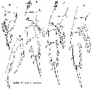 Issued from : P.A. Koeller & J.L. Littlepage in J. Fish. Red. Board Can., 1976, 33. [p.1551, Fig.3]. Female: A-D, P1 to P4, respectively. Male: E, P5. Nota: P1 similar to female with the following exception: long, fine spinules present on external margin of coxa. Remaining swimming legs with segmentation and spinulation as in female. P5 with coxa of left leg about as long as both basipods (= coxa and basis) of right leg combined; basis of left leg slightly shorter than coxa. Exopods of both legs 3-segmented, endopods 1-segmented, more or less rudimentary. Both rami of left leg longer than those of right leg. Right endopod with weak terminal seta. Last segment of left exopod and distal part of 2nd segment with fine spinules on the inner margin. Terminal segment of right exopod with short terminal seta and long slender spine about 1.3 times length of segment.
|
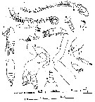 Issued from : P.A. Koeller & J.L. Littlepage in J. Fish. Red. Board Can., 1976, 33. [p.1550, Fig.2]. Male: A, right A1; B, left A1; C, Mx1; D, posterior part of body (lateral); E, Mx2; F, habitus (lateral); G, Md; H, Mxp; I, A2; J, habitus (dorsal); K, posterior part of body (dorsal). Nota: - Left A1 22-segmented; right A1 21-segmented. 8th and 10th segments of both antennae fused. 8th segment formed from 3 segments with faint partial suture line between 2nd and 3rd of these segments. 10th segment formed from 2 segments with partial suture line between them. Aesthetascs damaged, but probably 2 or more present on each proximal segment up to and including the 8th. 1 distal aesthetasc on all other segments. Right A1 with last 6 segments considerably enlarged. Distal portion bent backward at 3rd and 4th segment from end and curled slightly inward. Each distal seta of terminal segments developed into stout spine with point drawn out into fine hair. Otherwise similar to left A1. - A2 similar to female except differences in setation: 2nd endopodal segment without setae; terminal segment of exopod with reduced number of setae. - Mouth parts reduced. - Md without masticatory edge. Mx1 without setae on inner lobes. - Mx2 particularly small, lobes distinguishable, setation reduced. - Mxp more slender than in female; 2nd basal segment with 1 median seta. 1st endopodal segment with 4 setae, 2nd and 3rd endopodal segments with 3 setae each, 4th and 5th endopodal segments with 4 setae each.
| | | | | Compl. Ref.: | | | Bradford-Grieve, 2004 (p.283); Galbraith, 2009 (pers. comm.) | | | | NZ: | 1 | | |
|
Distribution map of Azygokeras columbiae by geographical zones
|
| | | | Loc: | | | NE Pacif. (British Columbia: Bute Inlet, Vancouver Is.)
Type locality: 50°27'N, 125°04'W. | | | | N: | 2 | | | | Lg.: | | | (209) F: 2,84-2,58; M: 2,1-2,08; {F: 2,58-2,84; M: 2,08-2,1} | | | | Rem.: | hyperbenthic (till 500 m);
Koeller & Littlepage (1976, p.1551) point out that asymmetry of A1 male does not involve an articulation as in Heterarthrandia. The right A1 appears to be used as a grasping organ as evidenced by the enlarged, curled distal portion, and by the stout spines directed toward the center of the curl. | | | Last update : 02/03/2015 | |
|
|
 Any use of this site for a publication will be mentioned with the following reference : Any use of this site for a publication will be mentioned with the following reference :
Razouls C., Desreumaux N., Kouwenberg J. and de Bovée F., 2005-2026. - Biodiversity of Marine Planktonic Copepods (morphology, geographical distribution and biological data). Sorbonne University, CNRS. Available at http://copepodes.obs-banyuls.fr/en [Accessed February 20, 2026] © copyright 2005-2026 Sorbonne University, CNRS
|
|
 |
 |


