|
|
 |
Fiche d'espèce de Copépode |
|
|
Calanoida ( Ordre ) |
|
|
|
Clausocalanoidea ( Superfamille ) |
|
|
|
Phaennidae ( Famille ) |
|
|
|
Xanthocalanus ( Genre ) |
|
|
| |
Xanthocalanus medius Tanaka, 1937 (F) | |
| | | | | | | Syn.: | X.anthocalanus media : Brodsky, 1950 (1967) (p.232, figs.F) | | | | Ref.: | | | Tanaka, 1937 (p.258, Descr.F, figs.F); Tanaka, 1960 a (p.96, figs.F); Bradford & al., 1983 (p.71) | 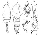 issued from : O. Tanaka in Publ. Sero Mar. Biol. Lab., 1960, VIII (1). [p.97, Fig.87]. Female (Suruga Bay, Japan): a, habitus (dorsal); b, forehead (lateral); c, last thoracic segment and urosome (lateral right side); d, rostrum (anterior aspect); e, distal joints of Mx2; f, P2; g, P5. Nota: Head and 1st thoracic segment separate, 4th and 5th fused. Rostrum bifurcate without any slender filaments at the apex. Lateral distal margin of the last thoracic segment produced triangularly with a point at the apex, when viewed from the dorsal. Cephalothorax 2.55 mm, abdomen 0.75 mm. The abdominal segments and caudal rami in the proportional lengths 35 : 25 : 19 : 2 : 19 = 100. The first 3 abdominal segments covered with scattered spinules; distal margin furnished with fine teeth. Anal segment haired on the dorsal as well as on the ventral surface. The genital segment bears on the middorsal line a rounded lamellous plate and a slender spine (when viewed from the lateral). A1 24 segmented, extends to the distal margin of the genital segment; aesthetasks on the joint 2, 3, 5, 8, 12, 14 and 19. A2 exopodite a little longer than the endopodite. Mx2 has 8 sensory setae on the endopod (of which 7 are bud-like and one is long vermiform). P5 resembles that of X. pinguis; the line of demarcation between the 2nd and 3rd segments undetectable; posterior surface of segments furnished with spinules; inner margin of the 2nd segment with 2 groups of poor denticles.
|
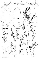 Issued from : O. Tanaka in Japanese J. Zool., 1937, VII, 13. [Pl. XVII]. As Xanthocalanus media. Female (from coast of Heda, Japan): 1, habitus (dorsal); 2, head (lateral); 3, abdomen (lateral); 4, rostrum; 5, A1; 6, distal part of Mx2; 7, Mxp; 8, P1; 9, P2; 10, P5. P1 and P5 at the same scale. Nota: Abdomen 4-segmented; comparative length of abdomen and caudal rami 35 : 25 : 19 : 2 : 19 = 100. Md: manducatory part long and slender with rather weak teeth; basis with 3 long spines; exopodite and endopodite subequal in length, endopodite 2-segmented of which the proximal bears 2 setae and the distal 8. Mx1: outer lobe with 7 long and 2 shorter setae; inner lobe with 10 posterior and 4 anterior setae; 2nd basal segment with 4 anterior and 1 posterior setae; endopodite with 3+3+4 setae; exopodite with 10 setae. Mx2 slightly produced posteriorly; lobe 1 with 5, lobe 2 with 3, lobe 3 with 2 setae and 1 vermiform filament, lobe 4 besides 1 usual seta a delicate bristle without spinules and a strong, slightly curved and serrated spine, lobe 5 with a long usual seta and 2 short setae of which one is posteriorly and naked; the strong claw-like seta is more coarsely serrated than in the lobe 4; endopodite with 7 longer or shorter brush-shaped sensory setae as well as a long slender vermiform one. Comparative length of the 3 main divisions of Mxp as 47 : 45: 32, respectively; 1st basipodite with besides usual spines, 1 short brush-shaped sensory seta; 2nd basipodite about 3 times as long as wide; 2nd exopodal segment the longest.
P5 resembles that of Xanthocalanus pinguis Farran.
|
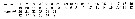 Issued from : O. Tanaka in Japanese J. Zool., 1937, VII, 13. [p.258]. As Xanthocalanus media. Female: Length of the segments on A1. Nota: A1 24-segmented, extend to the distal end of the genital segment. Large aesthetascks on the segment 2, 3, 5, 8-9, 12, 14, 19.
| | | | | Ref. compl.: | | | Sewell, 1948 (p.560) | | | | NZ: | 2 | | |
|
Carte de distribution de Xanthocalanus medius par zones géographiques
|
| | | | Loc: | | | Japan (Suruga) | | | | N: | 2 | | | | Lg.: | | | (128) F: 3,3; {F: 3,30} | | | | Rem.: | In the vertical haul 500-250 m. | | | Dernière mise à jour : 02/02/2015 | |
|
|
 Toute utilisation de ce site pour une publication sera mentionnée avec la référence suivante : Toute utilisation de ce site pour une publication sera mentionnée avec la référence suivante :
Razouls C., Desreumaux N., Kouwenberg J. et de Bovée F., 2005-2026. - Biodiversité des Copépodes planctoniques marins (morphologie, répartition géographique et données biologiques). Sorbonne Université, CNRS. Disponible sur http://copepodes.obs-banyuls.fr [Accédé le 08 avril 2026] © copyright 2005-2026 Sorbonne Université, CNRS
|
|
|
|


;)
;)
