|
|
 |
Fiche d'espèce de Copépode |
|
|
Calanoida ( Ordre ) |
|
|
|
Clausocalanoidea ( Superfamille ) |
|
|
|
Phaennidae ( Famille ) |
|
|
|
Xanthocalanus ( Genre ) |
|
|
| |
Xanthocalanus penicillatus Tanaka, 1960 (F) | |
| | | | | | | Syn.: | ? Xanthocalanus sp. Bradford, 1972 (p.40, figs.F) | | | | Ref.: | | | Tanaka, 1960 a (p.94, Descr.F, figs.F); Chen & Zhang, 1965 (p.58, 125: Rem.); ? Bradford & al., 1983 (p.71, figs.F, Rem.); Chihara & Murano, 1997 (p.855, Pl.142: F); Bradford-Grieve, 2004 (p.635: Rem.) | 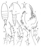 issued from : O. Tanaka in Publ. Sero Mar. Biol. Lab., 1960, VIII (1). [p.95, Fig.86]. Female (from Suruga Bay, Japan): a, habitus (dorsal); b, forehead (lateral); c, last thoracic segment and urosome (lateral left side); d, rostrum (anterior aspect); e, endopod of Mx1; f, Mx2; g, Mxp; h, P1; i, P2; j, endopod of P3; k, P4; l, P5. Nota: Head and 1st thoracic segment separate, 4th and 5th fused. Lateral distal corner of the last thoracic segment produced triangularly with a point at the apex. Rostrum ends into 2 filaments; basal part slightly swollen in lateral aspect. Cephalothorax 2.66 mm; abdomen 0.87 mm. The abdominal segments and furca in the proportional lengths 37 : 29 : 16 : 8 : 10 = 100. Genital segment as long as wide, inflated toward the distal end. The first 3 abdominal segments furnished with scattered spinules, the distal margin of the segments smooth. Caudal rami as long as wide. A2 exopodite slightly longer than the endopodite (22 : 20); the distal segment of endopodite with 6 + 6 setae. Md exopodite as long as the endopodite. Mx1 and Mx2 as in X. pinguis. Mx1 with 9 setae on the outer lobe; 10 setae on the exopodite; endopodite with 5+3+3 setae; 2nd basal with 5 setae; 3rd inner lobe with 4 setae, 2nd inner lobe with 5 setae, 1st inner lobe with 13 setae. Mx2carries both bud-like and vermiform setae on the endopodite. Mxp basal segments of about bequal lengths; segments of endopodite short. P5 3-sgmented; 3rd segment narrow in the distal half, with 4 spines, of which the inner marginal one the longest, the segment has a longitudinal row of spinules on the posterior surface; 2nd segment with groups of spines on the posterior as well as the anterior surface; 1st segment with denticles on the inner distal margin. Remarks: The specimen resembles closely X. profundus Sars, but is distinguished from it by small in size; complete fusion of the 4th and 5th thoracic segments; scattered spinules on the 1st to 3rd abdominal segments; the shape of P5.
|
 issued from : O. Tanaka in Publ. Sero Mar. Biol. Lab., 1960, VIII (1). [p.94]. Female: Proportions of segments of A1. Nota: A1 24-segmented, extends to the distal margin of the 5th thoracic segment.
|
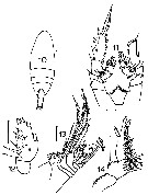 issued from : J.M. Bradford in Mem. N. Z. Oceonogr. Inst., 1972, 54. [p.42, Fig.9, (10-14)]. Xanthocalanus sp. Synonymy doubtful. Female (from Kaikoura, New Zealand): 10, habitus (dorsal); 11, P3; 12, endopod of P2; 13, Mx2; 14, P5. Scale bars: 1 mm (10); 0.1 mm (11, 12, 13, 14). Nota: P3 with 2-segmented exopod and 3-segmented endopod on one side whereas on the other side there are 3 segments in both rami. P4 with 2-segmented exopod and endopod on one side and on the other side are 3-segmented. The unequal development of these legs suggests that this specimen could be deformed. This specimen resembles X. squamatus Farran, 1936, but the Mx2 are not alike.
|
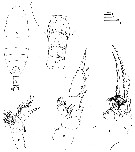 Issued from : J.M. Bradford, L. Haakonssen & J.B. Jillett in N. Z. Oceanogr. Inst. Memoir 90, 1983 [p.72, Fig.39]. Female (from 41°47'S, 175°01'E): A, habitus (dorsal); B, urosome (ventral); C, Mx2; D, P2; E, P5. Nota: Mx2 endopod with 1 worm-like and 7 brush-like sensory filaments, one of which is much thicker and shorter than others. P5 3-segmented, distal segment with 4 terminal spines. All three segments have additional small spines and spinules. Presence of small spines on the urosome.
| | | | | NZ: | 1 + 1 douteuse | | |
|
Carte de distribution de Xanthocalanus penicillatus par zones géographiques
|
| | | | Loc: | | | Japan (Suruga), ? New Zealand (Kaikoura) | | | | N: | 2 | | | | Lg.: | | | (4) F: 3,53; (9) F: 3,4; (116) F: 3,41; {F: 3,40-3,53} | | | | Rem.: | Bradfod-Grieve (2004) suppose que la forme citée en 1983 n'appartient pas à cette espèce , mais pourrait être X. stewarti. | | | Dernière mise à jour : 14/04/2015 | |
|
|
 Toute utilisation de ce site pour une publication sera mentionnée avec la référence suivante : Toute utilisation de ce site pour une publication sera mentionnée avec la référence suivante :
Razouls C., Desreumaux N., Kouwenberg J. et de Bovée F., 2005-2025. - Biodiversité des Copépodes planctoniques marins (morphologie, répartition géographique et données biologiques). Sorbonne Université, CNRS. Disponible sur http://copepodes.obs-banyuls.fr [Accédé le 03 décembre 2025] © copyright 2005-2025 Sorbonne Université, CNRS
|
|
|
|




;)
;)
;)
;)