|
|
 |
Fiche d'espèce de Copépode |
|
|
Calanoida ( Ordre ) |
|
|
|
Diaptomoidea ( Superfamille ) |
|
|
|
Pontellidae ( Famille ) |
|
|
|
Calanopia ( Genre ) |
|
|
| |
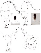
Calanopia aurivilli Cleve, 1901 (F,M) | |
| | | | | | | Syn.: | Calanopia aurivillei : Rose, 1956 (p.461) | | | | Ref.: | | | Cleve, 1901 (p.37, figs.F); Thompson & Scott, 1903 (p.235, 251); A. Scott, 1909 (p.181, figs.F,M); Sewell, 1912 (part. p.368); 1914 a (p.232, Rem.); Früchtl, 1924 b (p.57); Farran, 1929 (p.210, 273); Sewell, 1932 (p.341); Farran, 1936 a (p.115); Kasturirangan, 1963 (p.47, 48, figs.F,M); Saraswathy, 1966 (1967) (p.85); Silas & Pillai, 1976 (p.784, figs.F,M, Rem.); Bradford-Grieve, 1999 b (p.183, figs.F,M, figs.184, 194); Mulyadi, 2002 (p.36, figs.F,M, Rem.); Othman & Toda, 2006 (p.306, F,M); Phukham, 2008 (p.73, figs.F,M); Lacuna & al., 2013 (p.64, figs.F,M, Rem.: p.77, morphological variation) | 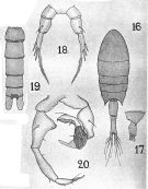 issued from : A. Scott in Siboga-Expedition, 1909, XIX a. [Plate XLVIII, Figs.16-20]. Female (from off Galle, Ceylon): 16, habitus (dorsal); 17, last thoracic and genital segments (left side)18, P5. Male: 19, urosome (dorsal); 20, P5.
|
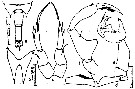 issued from : E.G. Silas & P.P. Pillai in J. mar. biol. Ass. India, 1973 (1976), 15 (2). [p.784, Fig.2]. Female (from Indian Ocean): a, urosome (dorsal); b, rostrum (anterior view); c, P5. Male: d, right A1 (geniculate); e, P5. Scale as in Calanopia minor.
|
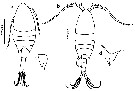
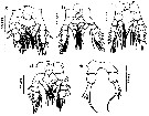
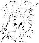 issued from : Mulyadi in Treubia, 2002, 32; [p.38, Fig.9]. Female (from Indonesian waters): a, habitus (dorsal); b, cephalon (lateral); c, metasomal somite 5 and urosome (lateral); d, rostrum (anterior view); e, P2; f, P3; g, P5. Male: h, habitus (dorsal); i, geniculate region of right A1; j, P5.
|
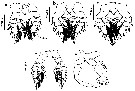
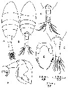 issued from : B.H.R. Othman & T. Toda in Coastal Mar. Sc., 2006, 30 (1). [p.307, Fig.2]. Female (from Sister's Island, Singapore): A, habitus (dorsal); D, posterior part of prosome and urosome (dorsal); E, P5. Male: B, habitus (dorsal); C, posterior part of prosome and urosome (dorsal); F, P5. Nota Female: Prosome to urosome length ratio 2.04 : 1. - Cephalon without lateral hook. - Prosome produced into posteriorly directed acute process. - Urosome 2-segmented. - Genital segment shorter than anal segment. - P5 symmetrical and uniramous; exopod 1-segmented apex terminates in 3 spines, inner being distinctly longer and plumose at its distal margin. Nota Male: Prosome to urosome length ratio 1.99 : 1. - Body similar to female. - Cephalon wiithout hook. - Right A1 geniculate. - Urosome 5-segmented, naked. - P5 asymmetrical and chelate; right leg 4-segmented, proximal inner margin of basis swollen, exopodal segment 1 with well developed thumb, claw spoo-shapede, slightly swollen at tip with 1 outer marginal seta, 1 terminal and 2 inner marginal; left leg basis swollen and gibbose, exopodal segment 1 with distolateral seta, terminal segment with 2 unequal apical spines.
|
 issued from : N. Phukham in Species diversity of calanoid copepods in Thai waters, Andaman Sea (Master of Science, Univ. Bangkok). 2008. [p.155, Fig.29]. Female (from W Malay Peninsula): a, habitus (dorsal); b, urosome; c, P5. Male: d, habitus (dorsal); e, P5. Body length after the drawings: F = 1.114 mm; M = 1.129 mm.
|
 ssued from : M.L.D.G. Lacuna, D.C. Sagrado, R.O. Mejorada, D.D. Simyunn & M.J.J. Pueblos in ABAH Bioflux, 2013, 5 (1). [p.65, Fig.13]. Female (from Iligan Bay, Mindanao): a, habitus (dorsal); c, rostrum. Male: b, habitus (dorsal); d, rostrum. Nota: The posterior corners of the last metasomal segment for both sexes are drawn out into spines.
|
 ssued from : M.L.D.G. Lacuna, D.C. Sagrado, R.O. Mejorada, D.D. Simyunn & M.J.J. Pueblos in ABAH Bioflux, 2013, 5 (1). [p.65, Fig.14]. Female: a, urosome (ventral). Male: b, urosome (dorsal).
|
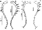 ssued from : M.L.D.G. Lacuna, D.C. Sagrado, R.O. Mejorada, D.D. Simyunn & M.J.J. Pueblos in ABAH Bioflux, 2013, 5 (1). [p.66, Figs.15, 16]. Female: a, left A1; b, right A1. Male: c, left A1; d, right A1. Nota: Both A1 female 17-segmented. For the male, left A1 17-segmented, while the right A1 is 12-segmented, geniculation at segments 7 and 8. The antennules of both sexes are terminated with 2 setae, each being distributed at the inner and outer margins.
|
 ssued from : M.L.D.G. Lacuna, D.C. Sagrado, R.O. Mejorada, D.D. Simyunn & M.J.J. Pueblos in ABAH Bioflux, 2013, 5 (1). [p.67, Fig.17]. Female: a, P1; b, P2; c, P3; d, P4, e, P5.
|
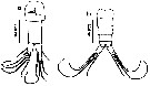
 ssued from : M.L.D.G. Lacuna, D.C. Sagrado, R.O. Mejorada, D.D. Simyunn & M.J.J. Pueblos in ABAH Bioflux, 2013, 5 (1). [p.67, Fig.18]. Male: a, P1; b, P2; c, P3; d, P4; e, P5.
|
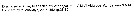 Calanopia aurivilli Calanopia aurivilli. Female: 1 - P5 exopod 1-segmented. 2 - Exopod of P5 with 4 spines; 3 - Exopod of P5 with 3 small spines and 1 long spine; 4 - Exopodal segment of P5 with 3 subequal small lateral spines and 1 long medial spine (longer than segment itself). Male: 1 Left P5 longer than right one ; basis of left P5 swollen proximally. 2 2nd exopodal segment of right P5 nearly 2/5 length of 1st exopodal segment ; coxa of right P5 about or less than 1.4 times as long as basis. 3 Basis of left P5 swollen proximally without any spines or processes.
| | | | | Ref. compl.: | | | Sewell, 1948 (p.323); C.B. Wilson, 1950 (p.174); Krishnaswamy, 1953 (p.138); Patel, 1975 (p.660); Carter, 1977 (1978) (p.36); Madhupratap & Haridas, 1986 (p.105, tab.1); Othman & al., 1990 (p.561, 564, Table 1); McKinnon, 1991 (p.471); Mauchline, 1998 (tab.8); Jitlang & al., 2008 (p.65, Table 1); Fernandes, 2008 (p.465, Tabl.2); McKinnon & al., 2008 (p.844: Tab.1); Cornils & al., 2010 (p.2076, Table 3); Shanthi & Ramanibai, 2011 (p.132, Table 1); Maiphae & Sa-ardrit, 2011 (p.641, Table 2, 3, Rem.); Jagadeesan & al., 2013 (p.27, Table 3, seasonal abundance); | | | | NZ: | 4 | | |
|
Carte de distribution de Calanopia aurivilli par zones géographiques
|
| | | | | | | | | | Loc: | | | SE South Africa (off Durban, Natal), NW India (Saurashtra coast), Trivandrum coast, Sri Lanka, Madras, G. of Mannar, Bay of Bengal, Burman coast, Andaman Sea, W Malay Peninsula, Strait of Malacca (Singapore), G. of Thailand, Indonnesia (S Java, Semau Sound, SE Celebes Sea, Philippines, Mindanao (Iligan Bay), Viet-Nam (Cauda Bay), off Hong Kong, Australia (G. of Carpentaria, Noth West Cape, Great Barrier), off New Zealand (NW North Island)
Type locality: Semau Sound (southwestern Timor). | | | | N: | 23 | | | | Lg.: | | | (5) F: 1,34; M: 1,12; (34) F: 1,32-1,27; M: 1,18-1,17; (35) F: 1,45; M: 1,38; (334) F: 1,34; M: 1,12; (530) F: 1,2; M: 1,1; (795) F: 1,3; M: 1,1; (1086) F: 1,05-1,20; M: 0,98-1,08; (1087) F: 1,2-1,25; M: 1,05-1,1; (1130) F: 1,56; M: 1,50; {F: 1,05-1,56; M: 0,98-1,50} | | | | Rem.: | épipélagique.
Voir aussi les remarques en anglais | | | Dernière mise à jour : 03/12/2020 | |
|
|
 Toute utilisation de ce site pour une publication sera mentionnée avec la référence suivante : Toute utilisation de ce site pour une publication sera mentionnée avec la référence suivante :
Razouls C., Desreumaux N., Kouwenberg J. et de Bovée F., 2005-2026. - Biodiversité des Copépodes planctoniques marins (morphologie, répartition géographique et données biologiques). Sorbonne Université, CNRS. Disponible sur http://copepodes.obs-banyuls.fr [Accédé le 31 mars 2026] © copyright 2005-2026 Sorbonne Université, CNRS
|
|
|
|


;)
;)
;)
;)
;)
;)
;)
;)
;)
;)

