|
|
 |
Fiche d'espèce de Copépode |
|
|
Calanoida ( Ordre ) |
|
|
|
Diaptomoidea ( Superfamille ) |
|
|
|
Pontellidae ( Famille ) |
|
|
|
Epilabidocera ( Genre ) |
|
|
| |
Epilabidocera longipedata (Sato, 1913) (F,M) | |
| | | | | | | Syn.: | Pontella longipedata Sato, 1913 (p.41, figs.); Marukawa, 1927 (p.1223, figs.); Mori, 1937 (1964) (p.96, figs.M); Sewell, 1948 (p.408); Kim & al., 1993 (p.270);
Pontella pulvinata C.B. Wilson, 1950 (p.295, Descr. F, M, figs.F,M); Marukawa & Tanaka, 1965 (p.476, fig.); Yamazi, 1966 (p.231, figs.).
? Paralabidocera amphitrites McMurrich, 1916; Willey, 1920 a (p.17, figs.F,M]; Esterly, 1924 (p.99, figs.F,M, Rem.);
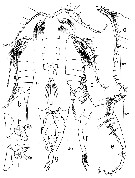
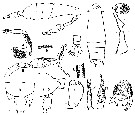
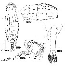
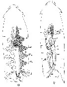
? Epilabidocera amphitrites : Davis, 1949 (p.64, figs.F,M); Brodsky, 1950 (1967) (p.413, figs.F,M); Ponomareva, 1961 (p.103); Park, 1966 a (p.129, figs.F,M, Rem.: external and internal structures); Ikeda, 1976 (p.51, respiration rate); Peterson & Miller, 1976 (p.14, Table 1, 2, 3, abundance vs interannual variations); Gibson & Grice, 1977 (p.85, Table 1, copper pollution); Peterson & Miller, 1977 (p.717, Table 1, seasonal occurrence); Grice & Marcus, 1981 (p.125, dormant eggs, Rem.: p.135); Marcus, 1990 (p.413, tab.1, 2, fig.2: egg); Mauchline, 1998 (tab.40, 58); Pinchuk & Paul, 2000 (p.4, table 1, % occurrence); ; Hooff & Peterson, 2006 (p.2610); Hopcroft & al., 2009 (p.9, Table 3); Matsuno & al., 2011 (p.1349, Table 1, abundance vs years); DiBacco & al., 2012 (p.483, Table S1, ballast water transport); Takahashi M. & al., 2012 (p.393, Table 2, water type index); in CalCOFI regional list (MDO, Nov. 2013 ; M. Ohman, comm. pers.); Ohashi & al., 2013 (p.44, Table 1, Rem.); Questel & al., 2013 (p.23, Table 3, interannual abundance & biomass, 2008-2010); Eisner & al., 2013 (p.87, Table 3: abundance vs water structure); Coyle & al., 2014 (p.97, table 3) | | | | Ref.: | | | Heinrich, 1969 (p.1457, fig.1); Nishimura, 1969 (p.381, Rem.); Silas & Pillai, 1976 (p.773); Gardner & Szabo, 1982 (p.410, figs.F, M); Ohtsuka & al., 1996 b (p.153, figs.F: mouth parts, gut contents); Chihara & Murano, 1997 (p.871, Pl.145: F,M); Gomez-Gutiérrez & Peterson, 1999 (p.637) | 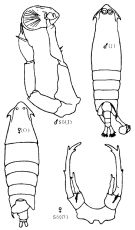 Issued from : K.A. Brodskii in Calanoida of the Far Eastern Seas and Polar Basin of the USSR. Opred. Fauna SSSR, 1950, 35 (Israel Program for Scientific Translations, Jerusalem, 1967) [p.413, Fig.293]. As Epilabidocera amphitrites [= E. longipedata after Nishimura (1969, p.382).
Female (from Sea of Okhotsk): habitus (dorsal); S5, P5.
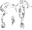
Male (from Sea of Japan): habitus (dorsal); S5, P5.
Nota Female:
- Head with characteristic lateral hook.
- Eyes present.
- Rostrum bifid, acute.
- Posterior thoracic segment with asymmetrical processes.
- Urosome composed of 3 segments.
- Genital segment with pterygoid process situated on left side and terminating posteriorly with long spine directed backward; ventral surface of segment with cariniform process.
- Urosomal segment 2 somewhat widened and elongate to the right in the distal part.
- Caudal rami symmetrical.
- A1 reaching end of thorax.
- P5 symmetrical, 2nd basal segment with 2 bristles on posterior surface; exopodite bearing 3 spines of unequal size on the distal end; endopodite with 2 spines.
Nota Male:
- Head with large lateral hooks.
- Ryes adjoining, larger than in female.
- Processes of terminal thoracic segment asymmetrical, right process thinner and much longer than the left and reaching middle of 3rd urosomal segment.
- 1st urosomal segment asymmetrical, expanded on right side, with small ventral process.
- Right A1 geniculate, with very swollen middle part.
- Chela of right leg of P5 round, comparatively small, with short thick, curved 'digits'; dorsal 'digit' bearing 2 spines. Distal segment of left leg bearing 2 apical and 1 lateral spine.
|
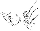
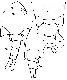 Issued from : A. Willey in Rep. Can. arct. Exped., 1920, VII: Crustacea, Part K: Marine Copepoda. [p.17K, Figs.14-17]. As Paralabidocera amphitrites.Female (from G. of Alaska): 14, forehead with eye and rostrum (lateral); 15, last thoracic segments and urosome with spermatophore (dorsal); 17, urosome (side view). Nota: Genital segment carries a large wing-like lobe on the left side, terminating in a recurved hook; this hook is easily lost, so that the lobe then appears to end bluntly. In side view the segment presents a ventral convexity and 2 small curved hooks, right and left of the genital opening. Male: 16, last thoracic segment and urosome (dorsal). Nota: Genital segment with a small lobe and notch on the left side
|
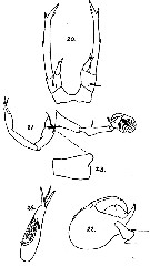 Issued from : A. Willey in Rep. Can. arct. Exped., 1920, VII: Crustacea, Part K: Marine Copepoda. [p.19K, Figs.20-24]. As Paralabidocera amphitrites. Female: 20, P5. Male: 21, P5; 22, chela of right leg of P5; 23, left basipod; 24, left terminal segment of P5.
|
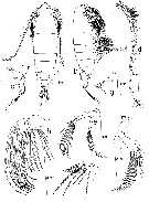 issued from : T.S. Park in La Cellule, 1966, LXVI (2). [p.135, Fig.1]. As Epilabidocera amphitrites. Female (from Friday Harbor, Washington): a, habitus (dorsal); b, idem (lateral view from right side); c, posterior portion of body (lateral view from left side); d, A1; e, A2; f, Md; g, Md (cutting edge of mandibular blade); h, Mx2; i, Mxp .
|
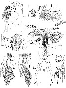 issued from : T.S. Park in La Cellule, 1966, LXVI (2). [p.137, Fig.2]. As Epilabidocera amphitrites. Female: a, head (with only maxillae; digestive tract shown in dotted outline; lateral view from left side); b, oral cone with Md (ventral); c, head (ventral); d, Mx1; e-f, P1-P2 (anterior aspects); g, P5 (anterior); h, urosome (ventral) laa = anterior lobe of labrum; lal = lateral lobe of labrum.
|
 issued from : T.S. Park in La Cellule, 1966, LXVI (2). [p.139, Fig.3]. As Epilabidocera amphitrites. Male: a, habitus (dorsal); b, idem (lateral view from left side); c, idem (lateral view from right side); d, left A1; e, right A1; f, left P5 (posterior); g, posterior portion of body with P5 (ventral); h, right P5; (anterior aspect); i, idem (posterior aspect).
|
 issued from : T. Mori in The pelagic Copepoda from the neighbouring waters of Japan, 1937 (2nd edit., 1964). [Pl.45, Figs.1-4]. As Pontella longipedata. Male (from Hokkaido in cold waters): 1, P5; 2, right A1; 3, habitus (dorsal); 4, P1.
|
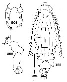
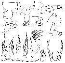 issued from : C.O. Esterly in Univ. Calif. Publs Zool., 1924, 26 (5). [p.100, Fig. J). As Paralabidocera amphitrites. Female (from San Francisco Bay): 1, posterior part of thorax and genital segment (dorsal); 2, posterior part of thorax and urosome (dorsal; from a second specimen); 3, idem (lateral); 4, postrior part of thorax and genital segment (lateral, left side; same specimen as figure 1); 5, forehead (lateral); 6, idem (ventral); 7, Mxp; 8, Md; 9, P1; 10, P2; 11, P3; 12, P4; 13, A2; 14, A1; 15, terminal part of exopod of A2; 16, P5. Nota: A1 24-segmented, extending to end of thorax. Abdomen 3-segmented, 1st segment always prolonged on left into wing projecting backwards and ending in sharp, spine-like process. (according to Willey, 5th segment of endopod with 3 setae, 4th 1, 3rd 1, 2nd 1, 1st 2, 2nd basal 3, distal lobe of 1st basal 1 very heavy, spinose bristle and 2 tiny setae. This is true for many specimens from bay but different individuals vary in number of bristles, as do appendages on two sides of same animal. jaw of Md with 5 teeth, ventral the largest.
|
 issued from : C.O. Esterly in Univ. Calif. Publs Zool., 1924, 26 (5). [p.87, Fig. B.). As Paralabidocera amphitrites. Male: 1, habitus (lateral); 2, cephalothorax and genital segment (dorsal); 3, right P5; 4, left P5; 5, some of the teeth on raised flange on thickened middle part of grasping A1; 6, right A1; 7, three posterior segments of abdomen and caudal rami; 8, some of the teeth on the segments proximal and distal to geniculation of grasping A1 (proximal teeth are at left); 9, posterior part of thorax and 1st and 2nd abdominal segments (dorsal); 10, posterior part of thorax, genital segment, basal segments of left P5 (lateral); 11, 2nd basal segment of left P5 (from side); 12-13, distal segment of left P5; 14, claw of right P5. Nota: Mxp 7-segmented
|
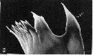 Issued from : S. Ohtsuka, M. Shimozu, A. Tanimura, M. Fukuchi, H. Hattori, H. Sasaki & O. Matsuda in Proc. NIPR Symp. Polar Biol., 1996, 9. [p.158, Fig.4, B]. Epilabidocera longipedata Female (from Bering Sea, Chukchi Sea): Mandibular cutting edge, ventralmost tooth (arrowed). Scale bar = 10 µm.
|
 Issued from : A. Willey in Rep. Can. arct. Exped., 1920, VII: Crustacea, Part K: Marine Copepoda. [p.18K, Figs.18, 19]. As Paralabidocera amphitrites. Male: 18, terminal portion of right A1. Nota: The right grasping A1 with the geniculation between the 18th and 19th segments, more like that of Pontella than that of Labidocera. 19, Mxp. Nota: Mxp 7-segmented, inner margin of basis denticulated; distally this segment carries 2 long setae and 1 additional shorter seta; Endopodal segment 1 with 2 setae, 2nd segment with 1, 3rd with 1, 4th with 1 and 5th with 3 apical setae. The distal lobe of coxa has 1 long seta and 2 very small anterior setae at its base, 1 of which was distinctly plumose. In the figure 19, one of the two setae of the middle lobe of coxa is lost, its place being indicated by a small mamelon.
|
 Issued from : C.B. Wilson in Bull. U.S. natn. Mus., 1950, 100 (14) (4). [Pl. 16, Figs. 198, 202, 203]. As Pontella pulvinata. Female (from Okhotsk Sea): 198, habitus (dorsal); 202, Md; 203, P5. Nota: Metasome 3 times as long as wide, strongly narrowed anteriorly. - Head bordered on each side by a wide membrane carrying a lateral hook. - 4th and 5th metasomal segments fused, posterior corners produced into thick fleshy triangular pads. These pads asymmetrical, the one on the right wider and longer than the left, nearly reaching the posterior margin of the genital segment; the posterior ends of the pads are broadly rounded with a minute spine at the tip (which is easily overlooked). - Urosome 3-segmented and very asymmetrical. - Dorsal surface of genital segment produced to the left and backward into a curved spine, which nearly reaches the tip of the caudal ramus. On the right side of segment and nearer the posterior margin is a short blunt process curved over ventrally, and usually concealed in dorsal view by the right pad at the corner of the thorax. On the left side is a rounded process projecting to the left and covered in dorsal view by the left pad at the corner of the metasome. - The basal abdominal segment is much larger than the anal segment, with an angular process at the center of the right side and a rounded process at the anterior corner of the left side.. - Anal segment about half as long as wide, as the basal segment, and its dorsal surface produced backward over the bases of the caudal rami in a 3-lobed process which reaches the center of the rami.; the latter are longer than wide and the left one is a little larger than the right. - A1 reach the center of the last thoracic segment and are very slender but moderately setose. - Exopod of A2 little shorter than endopod and considerably narrower. - Chewing blade of Md with a long conical tooth at the outer corner, then a shorter spherical tooth tipped with a spine, followed by 3 triangular teeth, the first two bifid at the tip. The palp has an exceptionally long basal portion and 2 short rami, each 1-segmented. - Endopod of P1 3-segmented, P2-P4 2-segmented. - Legs P5 slender, exopods twice as long as the endopods and each ramus 1-segmented. Endopods bifurcate at their tips, the inner branch longer than the outer. Each exopod with 3 spines at its tip, the middle one the longest, and a small spine on the outer margin near the center.
|
 Issued from : C.B. Wilson in Bull. U.S. natn. Mus., 1950, 100 (14) (4). [Pl. 16, Figs.199, 200, 201, 204]. As Pontella pulvinata. Male (from Okhotsk Sea): 199, habitus (dorsal); 200, middle segments of right A1; 201, urosome (ventral); 204, P5. Nota: Metasome proportionally narrower than in female. - Head with similar flanges that female, on each side armed with lateral hooks. - Posterior corners of last thoracic segment very asymmetrical, on the left side a pad similar to those in the female, on the right side a long and slender spine reaching back to the center of the antepenultimate segment of the abdomen. - Urosome 5-segmented. - Genital segment considerably enlarged and produced outward on the right side at the posterior corner into a short process cleft at its rip. - Urosomal segments 2 to 5 narrower than the genital segment, the first two segments of the same length, the third one 3/5, the anal segment 2/5 as long. - Caudal rami twice as long as wide and symmetrical. - A2, mouth parts, and P1 to P4 like those of the female.
- P5 each legs uniramose and 4-segmented. The terminal segment of the right leg transformed into a stout spherical chela without spines or processes. The terminal segment of the left leg is tipped with a slender, curved claw and a stout spine. The second segment of this leg carries at its distal end a small spine which might be regarded as the rudiment of an endopod.
|
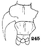 Issued from : C.B. Wilson in Bull. U.S. natn. Mus., 1950, 100 (14) (4). [Pl. 19, Fig.245]. As Pontella pulvinata. Female: 245, urosome (dorsal). Nota: Both pads of the posterior corners of metasome were removed for the drawing in figure 245.
|
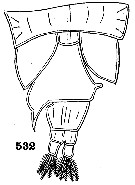 Issued from : C.B. Wilson in Bull. U.S. natn. Mus., 1950, 100 (14) (4). [Pl. 35, Fig.532]. As Pontella pulvinata. Female: 532, urosome (dorsal). Nota: The right pad of the posterior of the metasomal segment and the genital segment were separated under pressure, bringing the blunt process on the right side of the genital segment concealed in dorsal view.
|
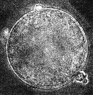 Issued from : N.H. Marcus in Mar. Biol., 1990, 105. [p.415, Fig.2, e]. Egg of Labidocera amphitrites from sediment (coast of northern California, in February-April 1989). Egg diameter (µm); 108.0 ± 3/0 (N = 15) (sediments, April); 110.4 ± 3.0. . Egg sueface smooth.
|
 Issued from : T.S. Park inLa Cellule, 1966, LXVI, 2. [p.185, Fig.14].Epilabidocera amphitrites. a, Female reproductive system (dorsal view); b, male reproductive system (dorsal view); c: canal; ca: coupling apparatus; de: ductus ejaculatorius; ga: genital aperture; gl: gland; jc: jelly coat of spermatophore; od: oviduct; oda: anterior diverticulum of oviduct; odl: lateral diverticuluim of oviduct; ov: ovary; sp: spermatophore; spa: spermatophore anlage; sps: spermatophore sac; st: spermatheca; sv: seminal vesicle; sz: spermatozoa; t: testes; tlv: transverse loop of vas deferens; tu: tubule; vc: vaginal cavity; vd: vas deferens.
| | | | | Ref. compl.: | | | Fleminger, 1967 a (tabl.1); Shih & al., 1971 (p.150); Peterson & Miller, 1975 (p.642, 650, Table 3, interannual abundance); Damkaer & Dey, 1982 (p.417, UV-B radiation effect); Mackas & Anderson, 1986 (p.115, Table 2); Dey & al., 1988 (p.321, 323, UV-B effect); Shih & Marhue, 1991 (tab.3); Marcus, 1996 (p.144); Madhupratap & al., 1996 (p.77, Table 2: resting eggs); Mauchline, 1998 (tab.40); Gomez-Gutiérrez & Peterson, 1999 (p.637, Table I, II, III, VI, figs.4, 5, 6, 7, abundance, egg production); Lenz & al., 2000 (p.338); Park, W & al., 2004 (p.464, tab.1); Choi & al., 2005 (p.710: Tab.III); Ohtsuka & al., 2008 (p.115, Table 5); Galbraith, 2009 (pers. comm.); Bollens & al., 2011 (p.1358, Table III) | | | | NZ: | 5 | | |
|
Carte de distribution de Epilabidocera longipedata par zones géographiques
|
| | |  Carte de 1996 Carte de 1996 | |
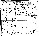 issued from : A.K. Heinrich in Zool. Zh., 1969, 48 (10) [p.1458, Fig.1]. issued from : A.K. Heinrich in Zool. Zh., 1969, 48 (10) [p.1458, Fig.1].
Distribution of two species of Pontellidae in the Pacific Ocean.
1: occurrence of Labidocera detruncata; 2: occurrence of Epilabidocera longipedata; 3: stations. |
| | | | Loc: | | | S Korea, Japan (Hokkaido), Japan Sea, La pérouse Straits, S & E Sakhalin, S. Kuril, Okhotsk Sea, Bering Sea (also eastern neritic), Chukchi Sea, off NW Alaska, Arct., G. of Alaska (Icy Strait), NE Alaska, Puget Sound, British Columbia, Fjord System (Alice Arm & Hastings Arm), Portland Inlet, Vancouver Island, Friday Harbor, Washington coast, Oregon (Yaquina Bay, off Newport), San Francisco Bay & Estuary, N California | | | | N: | 32 | | | | Lg.: | | | (22) F: 3,2-4; M: 2,3-3; (91) M: ±3,25; (280) F: 3,93-3,68; M: 3,28-2,78; (866) F: 3,2-4; M: 2,3-3,49; {F: 3,20-4,00; M: 2,30-3,49} | | | | Rem.: | Observé dans les ballasts des navires à San Francisco.
Voir aussi les remarques en anglais | | | Dernière mise à jour : 27/11/2020 | |
|
|
 Toute utilisation de ce site pour une publication sera mentionnée avec la référence suivante : Toute utilisation de ce site pour une publication sera mentionnée avec la référence suivante :
Razouls C., Desreumaux N., Kouwenberg J. et de Bovée F., 2005-2026. - Biodiversité des Copépodes planctoniques marins (morphologie, répartition géographique et données biologiques). Sorbonne Université, CNRS. Disponible sur http://copepodes.obs-banyuls.fr [Accédé le 15 juin 2026] © copyright 2005-2026 Sorbonne Université, CNRS
|
|
|
|


;)
;)
;)
;)
;)
;)
;)
;)
;)
;)
;)
;)
;)
;)
;)
;)
;)
;)
;)
