|
|
 |
Fiche d'espèce de Copépode |
|
|
Calanoida ( Ordre ) |
|
|
|
Diaptomoidea ( Superfamille ) |
|
|
|
Pontellidae ( Famille ) |
|
|
|
Labidocera ( Genre ) |
|
|
| |
Labidocera acuta (Dana, 1849) (F,M) | |
| | | | | | | Syn.: | Pontella acuta Dana,1849; Brady, 1883 (p.89, figs.F,M);
Labidocera acutum : Giesbrecht, 1892 (p.445, 458, 773, figs.F,M, no type 2: Pl.41, fig.29); Dakin & Colefax, 1940 (p.101, figs.F,M); De Decker, 1964 (p.15, 22, 28); De Decker & Mombeck, 1964 (p.13); Carter, 1977 (1978) (p.36); Dessier, 1983 (p.89, Tableau 1, Rem., %); | | | | Ref.: | | | Giesbrecht & Schmeil, 1898 (p.134, Rem. F,M); Thompson & Scott, 1903 (p.235, 251); A. Scott, 1909 (p.164, Rem.); Wolfenden, 1911 (p.361); Sewell, 1912 (p.353, 368, Rem.:M); Pesta, 1912 a (p.52, fig.F); 1913 (p.32); Sewell, 1914 a (p.232); Früchtl, 1924 b (p.55); Mori, 1929 (p.176, figs.F, M); Sewell, 1932 (p.351, figs. juv.); Dakin & Colefax, 1933 (p.206); Farran, 1936 a (p.116); Mori, 1937 (1964) (p.91, figs.F,M); Wilson, 1942 a (p.190, fig.F); Sewell, 1947 (p.248); C.B. Wilson, 1950 (p.241, figs.F,M); Chiba, 1956 (p.60, figs.F,M); Heinrich, 1960 a (p.43: fig.1); Brodsky, 1962 c (p.148, figs.F); Voronina, 1962 a (p.70, fig.2); Kasturirangan, 1963 (p.50, figs.F,M); Tanaka, 1964 c (p.254, Rem.F,M); Chen & Zhang, 1965 (p.98, figs.F,M); Saraswathy, 1966 (1967) (p.81); Silas & Pillai, 1967 (1969) (p.346, figs.F,M, Rem., biogéo.); Koga, 1968 (p.17, fig.: egg); Silas & Pillai, 1973 (1976) (p.795, figs.F,M, Rem.); Goswami & Goswami, 1974 (p.109, fig. caryotypes); Fleminger, 1975 (p.395); Marques, 1976 (p.995, fig.F); Greenwood, 1979 (p.95, figs.F,M, Rem.); Marques, 1982 (p.766); Kimmerer & al., 1985 (p.426); Ohtsuka & Onbé, 1991 (p.214); Chihara & Murano, 1997 (p.866, Pl.147,149: F,M); Barthélémy, 1999 a (p.9, Fig.7, F); Bradford-Grieve & al., 1999 (p.885, 959, figs.F,M); Bradford-Grieve, 1999 b (p.187, figs.F,M, Rem., figs.184, 194); Mulyadi, 2002 (p.49, figs.F,M, Rem.); Conway & al., 2003 (p.125, figs.F,M, Rem.); Othman & Toda, 2006 (p.308, figs.F,M); Phukham, 2008 (p.78, figs.F,M); Al-Yamani & al., 2011 (p.62, figs.F,M); Lacuna & al., 2013 (p.57, figs.F,M, Rem.); Sanu & al., 2016 (p.99, Table 1, 2, 3, molecular database); Abo-Taleb, 2019 (p.367, 369: Key F, M, figs.F, M).
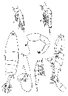
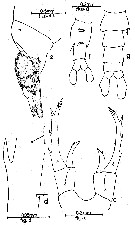
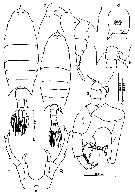
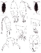
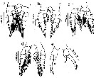
| 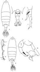 issued from: Q.-c Chen & S.-z. Zhang in Studia Marina Sinica, 1965, 7. [Pl.41, 5-10]. Female (friom E China Sea): 5, habitus (dorsal); 6, urosome (ventral); 7, right P5 (posterior). Male: 8, habitus (dorsal); 9, distal part of right A1; 10, P5 (posterior).
|
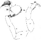
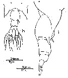
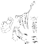
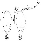
 issued from : J.G. Greenwood in Proc. R. Soc. Qd, 1979, 90. [p.96, Fig.1]. Female (from Moreton Bay, E Australia): a, forehead (lateral); b, urosome (dorsal); d, P5. Male: c, P5.
|
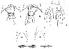
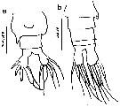
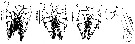
 issued from : E.G. Silas & P.P. Pillai in J. mar. biol. Ass. India, 1973 (1976), 15 (2). [p.796, Fig.9]. Female (from Indian Ocean): a, urosome (dorsal); b, 5th metasomal somite (lateral right side); c, forehead (lateral); d, P5. Male: e, urosome (dorsal); f, right A1 (geniculate part); g, P5; h-i, P5 (terminal segments, enlarged). Scale as in Calanopia minor.
|
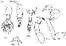
 Issued from : W. Giesbrecht in Systematik und Faunistik der Pelagischen Copepoden des Golfes von Neapel und der angrenzenden Meeres-Abschnitte. Fauna Flora Golf. Neapel, 1892, 19. Atlas von 54 Tafeln. [Taf.41, Figs.10, 20]. As Labidocera acutum. Female: 10, habitus (dorsal); 20, urosome (ventral).
|
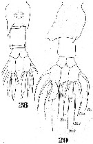 Issued from : W. Giesbrecht in Systematik und Faunistik der Pelagischen Copepoden des Golfes von Neapel und der angrenzenden Meeres-Abschnitte. Fauna Flora Golf. Neapel, 1892, 19. Atlas von 54 Tafeln. [Taf.41, Figs.28, 29]. As Labidocera acutum. Female: 28, urosome (ventral); 29, idem (dorsal). Nota: For Silas & P. Pillai (1969) the figure 29 belongs to the variant form: second type of Giesbrecht from the Arabian Sea and the Red Sea, and is a new species: L. pseudacuta Silas & Pillai, 1969
|
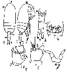
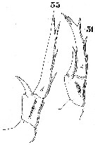 Issued from : W. Giesbrecht in Systematik und Faunistik der Pelagischen Copepoden des Golfes von Neapel und der angrenzenden Meeres-Abschnitte. Fauna Flora Golf. Neapel, 1892, 19. Atlas von 54 Tafeln. [Taf.25, Figs.31, 33]. As Labidocera acutum. Female: 31, P5; 33, P5 (variant).
|
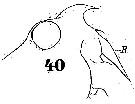 Issued from : W. Giesbrecht in Systematik und Faunistik der Pelagischen Copepoden des Golfes von Neapel und der angrenzenden Meeres-Abschnitte. Fauna Flora Golf. Neapel, 1892, 19. Atlas von 54 Tafeln. [Taf.41, Fig.40]. As Labidocera acutum. Male: 40, forehead (lateral).
|
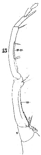 Issued from : W. Giesbrecht in Systematik und Faunistik der Pelagischen Copepoden des Golfes von Neapel und der angrenzenden Meeres-Abschnitte. Fauna Flora Golf. Neapel, 1892, 19. Atlas von 54 Tafeln. [Taf.23, Fig.15]. As Labidocera acutum. Male: 15, right A1 (distal part, posterior surface).
|
 Issued from : W. Giesbrecht in Systematik und Faunistik der Pelagischen Copepoden des Golfes von Neapel und der angrenzenden Meeres-Abschnitte. Fauna Flora Golf. Neapel, 1892, 19. Atlas von 54 Tafeln. [Taf.23, Figs.44, 46]. Male: 44, left P5 (anterior view); 46, right P5 (posterior view).
|
 Issued from : W. Giesbrecht in Systematik und Faunistik der Pelagischen Copepoden des Golfes von Neapel und der angrenzenden Meeres-Abschnitte. Fauna Flora Golf. Neapel, 1892, 19. Atlas von 54 Tafeln. [Taf.41, Fig.19]. As Labidocera acutum. Male: 19, 5th thoracic segment and urosome (dorsal).
|
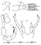
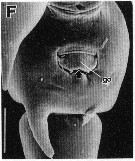
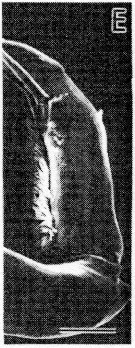 issued from : S. Ohtsuka & R. Huys in Hydrobiologia, 2001, 453/454. [p.452, Fig.7, E]. Male (from Japan): E, terminal exopodal segment of left P5. Scale bar = 0.5 mm.
|
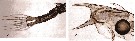
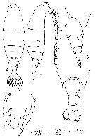
 issued from : T. Mori in The pelagic Copepoda from the neighbouring waters of Japan, 1937 (2nd edit., 1964). [Pl.41, Figs.1-5]. Female: 2, habitus (lateral, right side); 4, P5. Male: 1, habitus (dorsal); 3, forehead (lateral); 5, P5.
|
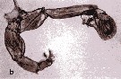
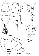
 issued from : T. Mori in Zool. Mag. Tokyo, 1929, 41 (486-487). [Pl. VII, Figs.5-10]. Female (from Chosen Strait, Korea-Japan): 5, P2; 6, P4. Male: 7, P4; 8, right A1; 9, habitus (dorsal); 10, P5.
|
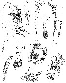
 issued from : E.G. Silas & P. Parameswaran Pillai J. Mar. biol. Ass. India, (1967) 1969, 9 (2). [p.350, Text-Fig.1, h, i].
Female: h, last thoracic segment and urosome (dorsal); i, same (lateral, right side).
|
 issued from : E.G. Silas & P. Parameswaran Pillai J. Mar. biol. Ass. India, (1967) 1969, 9 (2). [p.351, Text-Fig.2, f, j-k, o, n].
Female: f, caudal rami and cudal setae; j-k, urosome with spermatophore ( (dorsal and ventral, respectively); o, rostrum.
Male: n, rostrum.
|
 issued from : E.G. Silas & P. Parameswaran Pillai J. Mar. biol. Ass. India, (1967) 1969, 9 (2). [p.349].
Female: Proportions of segments of A1.
|
 issued from : E.G. Silas & P. Parameswaran Pillai J. Mar. biol. Ass. India, (1967) 1969, 9 (2). [p.356, Text-Fig.4, c-g].
Female: c, P5; d, part of exopod of left P5 enlarged; e, urosome with spermatophore (lateral); f, urosome (ventral).
Male: g, urosome (dorsal).
|
 issued from : E.G. Silas & P. Parameswaran Pillai J. Mar. biol. Ass. India, (1967) 1969, 9 (2). [p.355, Text-Fig.3, g-k].
Male: g, last thoracic esgment and urosome (dorsal); h, segments 16-23 of right A1 with parts of plates enlarged; i, P5; j-k, terminal part of left P5 (in two specimens).
|
 issued from : E.G. Silas & P. Parameswaran Pillai J. Mar. biol. Ass. India, (1967) 1969, 9 (2). [p.353].
Male: Proportions of the segments of the geniculate A1.
|
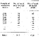 issued from : E.G. Silas & P. Parameswaran Pillai J. Mar. biol. Ass. India, (1967) 1969, 9 (2). [p.353].
Male: Frequency of occurrence of teeth on plates of geniculate A1.
|
 issued from : R.-M. Barthélémy in These Doct. Univ. Provence (Aix-Marseille I), 1999. [Fig.7, F]. Female (from Red Sea): F, external ventral view genital double-somite. go = genital operculum. Note the posterior bilobed margin. Scale bar: 0.050 mm.
|
 issued from : Y. Al-Yamani, V. Skryabin, A. Gubanova, S. khvorov & I. Prusova in Marine Zooplankton Prarctical Guide for the Northwestern Arabian Gulf, 2, 2011 [p.64, Fig.189, c-d]. Female (from NW Arabian Gulf): c, P5; d, last thoracic segment and urosome.
|
 issued from : Y. Al-Yamani, V. Skryabin, A. Gubanova, S. khvorov & I. Prusova in Marine Zooplankton Prarctical Guide for the Northwestern Arabian Gulf, 2, 2011 [p.65, Fig.190, c-d]. Male: c, urosome; d, forehead.
|
 issued from : Y. Al-Yamani, V. Skryabin, A. Gubanova, S. khvorov & I. Prusova in Marine Zooplankton Prarctical Guide for the Northwestern Arabian Gulf, 2, 2011 [p.65, Fig.190 b]. Male: b, P5.
|
 issued from : Mulyadi in Treubia, 2002, 32; [p.51, Fig.13]. Female (from Indonesian waters): a, habitus (dorsal); b, forehead (lateral); c, metasomal somite 5 and urosome (lateral); d, genital complex (ventral); e, P5. Male: f, habitus (dorsal); g-h, metasomal somite 5 and urosomal segments 1-2 (dorsal and right lateral); i, P5.
|
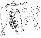
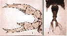
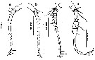
 Issued from : G.S. Brady in Rep. Scient. Results Voy. Challenger, Zool., 1883, 8 (23). [Pl. XXXVI]. Female: 1, habitus (right side); 6, Mx1; 7, Mxp; 9, P5; 11, posterior thoracic angles and urosome; 12, 2nd and 3rd setae of caudal ramus (highly magnified). Male: 2, left A1; 3, right A1; 4-5, portions of denticulated plates of geniculate portion of right A1; 8, P5; 10, posterior thoracic angles and urosome.
|
 issued from : B.H.R. Othman & T. Toda in Coastal Mar. Sc., 2006, 30 (1). [p.309, Fig.6]. Female (from Sister's Island, Singapore): A-B, habitus (dorsal and lateral, respectively); C-D, posterior part of prosome and urosome (lateral and dorsal, respectively); E, P5. Nota: Prosome to urosome length ratio 2.94 : 1. - Prosome ending in a single spiniform process. - Cephalon rounded with a median anterior hook, dorsal eye lenses moderate, separated. Lateral cephalic hook absent. - Thoracic process reaching beyond middle of genital segment. - Urosome 3-segmented. - Genital segment with a stout distolateral conical process on right margin. - P5 asymmetrical; right leg being stouter and longer than left, exopod with 3 outer, 1 inner, and 3 apical spines of which medial one longest; endopod bifurcated at apex.
|
 issued from : B.H.R. Othman & T. Toda in Coastal Mar. Sc., 2006, 30 (1). [p.309, Fig.7]. Male (from Sister's Island, Singapore): A-B, habitus (dorsal and lateral, respectively); C-D, posterior part of prosome and urosome (lateral and dorsal); E, P5. Nota: Prosome to urosome length ratio 2.96 : 1. - Body similar to female but anterior hook more pronounced, dorsal lenses slightly larger and closer together than female. - Thoracic process asymmetrical, left process similar to females, right produced into a curved process turned distolaterally and reaching distal end of urosomite 2. - Urosome 5-segmented. - Genital segment asymmerrtical, left side convex posteriorly, right side armed on posterior end with pointed process. - Right A1 geniculate, segment 17 naked, anterior margin of segment 18 with row of prominent denticles, extends proximally to almost whole length of segment 17, fused segments 19-21 with toothed plate extending to 2/3 length of the segment, segment 22 prolonged distally into spur-like process which is as long as its own segment. -P5 asymmetrical; right leg chelate, exopodal segment 1 orbicular, exopodal segment 2 short, broader medially, with 2 inner and 2 apical setae; left leg, exopodal segment 1 with distolateral spine, exopodal segment 2 ending in 3 finger-like processes, 1 small cresented basal process and 1 spine near distal end, inner margin of segment hirsute.
|
 issued from : N. Phukham in Species diversity of calanoid copepods in Thai waters, Andaman Sea (Master of Science, Univ. Bangkok). 2008. [p.159, Fig.33]. Female (from W Malay Peninsula): a, habitus (dorsal); b, same (lateral); c, posterior thoracic segment and urosome (oblque right lateral view); d, P5. Male: e, habitus (dorsal); f, urosome; g, P5. Body length after the drawings: F = 3.000 mm; M = 2.957 mm.
|
 Issued from : M.L.D.G. Lacuna, D.C. Sagrado, R.O. Mejorada, D.D. Simyunn & M.J.J. Pueblos in ABAH Bioflux, 2013, 5 (1). [p.58, Fig.2]. Female (from Sarangani Bay, Mindanao): a, habitus (dorsal). Male: b, habitus (dorsal). Nota: Male's eyes bigger than that of the female. The corners of posterior prosome are pointed, symmetrical in female and asymmetrical in male.
|
 ssued from : M.L.D.G. Lacuna, D.C. Sagrado, R.O. Mejorada, D.D. Simyunn & M.J.J. Pueblos in ABAH Bioflux, 2013, 5 (1). [p.59, Fig.3]. Female: a, urosome (ventral). Male: b, urosome (dorsal). Nota: Caudal ramus of both sexes with 5 thick setae of equal length
|
 ssued from : M.L.D.G. Lacuna, D.C. Sagrado, R.O. Mejorada, D.D. Simyunn & M.J.J. Pueblos in ABAH Bioflux, 2013, 5 (1). [p.559, Figs.4, 5]. Female: a, left A1; b, right A1. Male: c, left A1; d, right A1. Nota: A1 female symmetrical with 24 segments; A1 male asymmetrical with 22 segments.
|
 ssued from : M.L.D.G. Lacuna, D.C. Sagrado, R.O. Mejorada, D.D. Simyunn & M.J.J. Pueblos in ABAH Bioflux, 2013, 5 (1). [p.60, Fig.6]. Female: a, P1; b, P2; c, P3; d, P4; e, P5.
|
 ssued from : M.L.D.G. Lacuna, D.C. Sagrado, R.O. Mejorada, D.D. Simyunn & M.J.J. Pueblos in ABAH Bioflux, 2013, 5 (1). [p.60, Fig.7]. Male: a, P1; b, P2; c, P3; d, P5.
|
 Issued from : J.M. Bradford-Grieve, E.L. Markhaseva, C.E.F. Rocha & B. Abiahy in South Atlantic Zooplankton, edit. D. Boltovskoy. 1999, Vol. 2, Copepoda; [p.1067, Fig. 7.381]. Ce = cephalon; Ur = urosome; l = left leg; r = right leg,. B = basis; Exp1 = exopodal segment 1; Exp2-3 = exopodal segment 2-3; Exp3 = exopodal segment 3. Labidocera acuta. Female characters (from key, p.959): Posterior prosome corners directed posteriorly; urosomal segment 2 without any dorsal projections; anterior cephalosome with crest, visible in dorsal view as an anterior spine; cephalon without lateral hooks. Male characters (p.959): Right P5 basis long, more than twice length of exopodal segment 1.left P5 basis not process especially produced distally (compare with L. acutifrons).
|
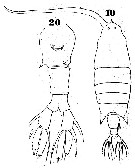
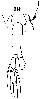
 Issued from : M.S. Kos in Field guide for plankton. Zool Institute USSR Acad., Vol. I, 1972. After Mori, 1937 (figs. 3, 11, 12) and Brodsky, 1962 (other figures). Female:: 1-2, habitus (dorsal and lateral (respectively); 3, forehead (dorsal); 4, same (lateral, left side); 5, abdomen (ventral); 6, same (lateral, right side); 7, corners of the last thoracic segment and abdomen (dorsal); 8, corner of the last thoracic segment and genital segment (lateral, left side); 9, P5. Male: 10, habitus (dorsal); 11, left P5; 12, right P5.
|
 Issued from : H.A. Abo-Taleb in Egyptian J. Aqua. Res., 2019, 45. [p.369]. Key to species of Labidocera in the Red Sea. Female: 1 - Anterior end of cephalosome with median spine-like crest in dorsal view. 1' - Other species without median crest on the anterior end of cephalosome. Male: 1 - Anterior end of cephalosome with a median spine-like crest in dorsal view. 1' - other species without a median crest on the anterior end of ceplalosome.
| | | | | Ref. compl.: | | | Cleve, 1904 a (p.191); Carl, 1907 (p.17); Sewell, 1948 (p.322); Krishnaswamy, 1953 (p.130); Chiba & al., 1957 (p.308); 1957 a (p.11); Yamazi, 1958 (p.151, Rem.); Heinrich, 1960 a (p.43: fig.1); Ganapati & Shanthakumari, 1962 (p.9, 15); Sherman, 1964 (p.481); Anraku & Azeta, 1965 (p.13, Table 2, fish predator); Fleminger, 1967 a (tabl.1); Delalo, 1968 (p.138); Heinrich, 1969 (p.1461, fig.3); Brodsky, 1972 (p.256); Kos, 1972 (Vol. I, figs.F,M, Rem.); Björnberg, 1973 (p.352, 387); Weikert, 1975 (p.137, chart); Patel, 1975 (p.660); Tranter, 1977 (p.598); Grice & Gibson, 1978 (p.23, tab.8); Chen Q-c, 1980 (p.795); Sreekumaran Nair & al., 1981 (p.493), Dessier, 1983 (p.89, Tableau 1 as acutum, Rem.: p.93, %); Guangshan & Honglin, 1984 (p.118, tab.); De Decker, 1984 (p.317, 352: chart); Brinton & al., 1986 (p.228, fig.11: spatial distribution, Table 1); Chen Y.-Q., 1986 (p.205, Table 1: abundance %, Table 2: vertical distribution); Madhupratap & Haridas, 1986 (p.105, 112: Rem., tab.1); Jimenez-Perez & Lara-Lara, 1988; Heinrich, 1988 (p.89, fig.2); Hernandez-Trujillo, 1989 (tab.1); 1989 a (tab.1); Cervantes-Duarte & Hernandez-Trujillo, 1989 (tab.3); Mizushima, 1990 (fig.2, 3, tab.2); Mitra & al., 1990 (fig.3); Othman & al., 1990 (p.561, 564, Table 1); Dai & al., 1991 (tab.1); Yoo, 1991 (tab.1); Godhantaraman, 1994 (tab.6); Hernandez-Trujillo, 1994 (tab.1); Shih & Young, 1995 (p.71); Kotani & al., 1996 (tab.2); Suarez-Morales & Gasca, 1997 (p.1525); Park & Choi, 1997 (Appendix); Sharaf & Al-Ghais, 1997 (tab.1); Ramaiah & Nair, 1997 (tab.1); Padmavati & al., 1998 (p.349); Achuthankutty & al., 1998 (p.1, Table 2, seasonal abundance vs monsoon); Hwang & al., 1998 (tab.II); Hsieh & Chiu, 1998 (tab.2); Wong & al, 1998 (tab.2); Suarez-Morales & Gasca, 1998 a (p.110); Smith S. & al., 1998 (p.2369, Table 6, moonsoon effects); Mauchline, 1998 (tab.8); Hernandez-Trujillo, 1999 (p.284, tab.1); El-Serehy, 1999 (p.172, Table 1, occurrence); Lavaniegos & Gonzalez-Navarro, 1999 (p.239, Appx.1); Fernandez-Alamo & al., 2000 (p.1139, Appendix); Suarez-Morales & al., 2000 (p.751, tab.1); Lo & al., 2001 (1139, tab.I); Hernandez-Trujillo & Suarez-Morales, 2002 (p.748, tab.1); Alvarez-Silva & al., 2003 (p.737, tab.2); Osore & al., 2003 (p.69); Hwang & al., 2003 (p.193, tab.2); Hsiao & al., 2004 (p.326, tab.1); Rezai & al., 2004 (p.489, tab.2); Lan & al., 2004 (p.332, tab.1); Lo & al.*, 2004 (p.218, fig.6); Lo & al., 2004 (p.89, tab.1); Kazmi, 2004 (p.228); Alvarez-Silva & al., 2005 (p.39); Rezai & al., 2005 (p.157, Table 5: spatial & temporal variations); Lavaniegos & Jiménez-Pérez, 2006 (p.146, tab.2, 3, Rem.); Lopez-Ibarra & Palomares-Garcia, 2006 (p.63, Tabl. 1, seasonal abundance vs El-Niño); Rakhesh & al., 2006 (p.93, Table 2, spatial distribution); Zuo & al., 2006 (p.163: tab.1); Hwang & al., 2006 (p.943, tabl. I); Hwang & al., 2007 (p.24); Rakhesh & al., 2008 (p.154, abundance vs stations); Morales-Ramirez & Suarez-Morales, 2008 (p.521); Ayon & al., 2008 (p.238, Table 4: Peruvian samples); Ohtsuka & al., 2008 (p.115, Table 5); Tseng L.-C. & al., 2008 (p.153, Table 2, occurrence vs geographic distribution); C.-Y. Lee & al., 2009 (p.151, Tab.2); Lan Y.-C. & al., 2009 (p.1, Table 2, % vs hydrogaphic conditions); Cornils & al., 2010 (p.2076, Table 3); Hernandez-Trujillo & al., 2010 (p.913, Table 2); W.-B. Chang & al., 2010 (p.735, Table 2, abundance); Shanthi & Ramanibai, 2011 (p.132, Table 1); Hsiao S.H. & al., 2011 (p.475, Appendix I); Kâ & Hwang, 2011 (p.155, Table 3: occurrence %); Maiphae & Sa-ardrit, 2011 (p.641, Table 2, 3, Rem.); Tutasi & al., 2011 (p.791, Table 2, abundance distribution vs La Niña event); Pillai H.U.K. & al., 2011 (p.239, Table 3, vertical distribution); Guo & al., 2011 (p.567, table 2, indicator); Lavaniegos & al., 2012 (p. 11, Table 1, seasonal abundance, Appendix); Tseng & al., 2012 (p.621, Table 3: abundance); Jose & al., 2012 (p.20, fig.3 a,b,c: % vs monsoon); Naz & al., 2012 (p.61, Table 4, relative abundance); Palomares-Garcia & al., 2013 (p.1009, Table I, abundance vs environmental factors); in CalCOFI regional list (MDO, Nov. 2013; M. Ohman, comm. pers.); Tseng & al., 2013 (p.507, seasonal abundance); Rakhesh & al., 2013 (p.7, Table 1, abundance vs stations); Jagadeesan & al., 2013 (p.27, Table 3, 5, fig.11, seasonal distribution); Anjusha & al., 2013 (p.40, Table 3, abundance & feeding behavior);Varadharajan & Soundarapandian, 2013 (p.2: occurrence vs stations); Mendoza Portillo, 2013 (p.37: Fig.7, seasonal dominance); Oh H-J. & al., 2013 (p.192, Table 1, occurrence); Hwang & al., 2014 (p.43, Appendix A: seasonal abundance); Beltran Castro, 2014 (p.39, Table 1, 2: molecular CO1); Jerez-Guerrero & al., 2017 (p.1046, Table 1: temporal occurrence); Palomares-Garcia & al., 2018 (p.178, fig.3: relative frequency, Table 1) | | | | NZ: | 15 | | |
|
Carte de distribution de Labidocera acuta par zones géographiques
|
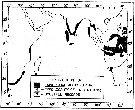
| | | | | | | | | | | |  Issued from : A.K. Heinrich in Trudy Inst. Okeanol., 1960, 41. [p.43, Fig.1]. Issued from : A.K. Heinrich in Trudy Inst. Okeanol., 1960, 41. [p.43, Fig.1].
Distribution in the equatorial and tropical West Pacific of seven species of Pontellidae (Cruise of the R/V 'Vityaz', November 1957 to December 1958.
1: Stations; 2: Pontellopsis villosa; 3: Pontella fera; 4: Labidocera acutifrons; 5: Pontella whiteleggi; 6: Pontella tenuiremis; 7: Pontella chierchiae; 8: Labidocera acuta. |
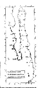
 issued from : E.G. Silas & P. Parameswaran Pillai J. Mar. biol. Ass. India, (1967) 1969, 9 (2). [p.361, Text-Fig.8]. issued from : E.G. Silas & P. Parameswaran Pillai J. Mar. biol. Ass. India, (1967) 1969, 9 (2). [p.361, Text-Fig.8].
Map showing the distribution of L. acuta in the Indian Ocean. It may be noted that most of the records are from coastel waters.
Nota: According to Fleminger (pers. comm.) the records from the Atlantic are based on misidentification. L. acuta is known at present from both neritic as well as island associated waters in the Pacific Ocean and the Indian Ocean. |
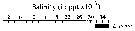 issued from : C.T. Achuthankutty, N. Ramaiah & G. Padmavati in Pelagic biogeography ICoPB II. Proc. 2nd Intern. Conf. Final report of SCOR/IOC working group 93, 9-14 July 1995. Workshop Report No. 142, Unesco, 1998. [p.8, Fig.6]. issued from : C.T. Achuthankutty, N. Ramaiah & G. Padmavati in Pelagic biogeography ICoPB II. Proc. 2nd Intern. Conf. Final report of SCOR/IOC working group 93, 9-14 July 1995. Workshop Report No. 142, Unesco, 1998. [p.8, Fig.6].
Salinity ranges for L. minuta in coastal and estuarine waters of Goa (India).
Shaded area indicates the range of higher abundance. |
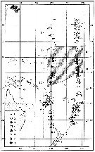
 Issued from : E. Brinton, A. Fleminger & D. Siegel-Causey in CalCOFI Rep., 1986, 27. [p.244, Fig.11]. Issued from : E. Brinton, A. Fleminger & D. Siegel-Causey in CalCOFI Rep., 1986, 27. [p.244, Fig.11].
labidocera copepods of the Gulf of California and Baja California. Three 'Labidocera species, not closely related, which mix with both ''L. jollae group'' and ''L. trispinosa group. All have more offshore habitat preferences: two are oceanic, L. acutifrons being broadly tropical, whereas L. detruncata is equatorial; L. acuta is broadly neritic and equatorial. |
| | | | Loc: | | | South Africa (E & W), G. of Guinea, Brazil (in Björnberg, 1965), Yucatan (in Suarez-Morales & Gasca (1997), Ireland (in Giesbrecht & Schmeil, 1898), Irish Sea (Man Is.), E Medit. (Port Saïd), Suez Canal, Red Sea, G. of Aden, G. of Oman, Arabian Gulf, UAE coast, Arabian Sea, Karachi coast, Laccadive Is., Maldive Is., Port Natal, off Natal, off SE Madagascar, Nosy Bé, Rodrigues Is., Seychelles, Sri Lanka, Indian, India (Saurashtra coast, Mangalore coast, Bombay, Goa, Gulf of Mannar, Palk Bay, Pointcalimere-Manamelkudi, Madras, Lawson's Bay, Mandarmani), E India (coastal, Godavari region, Kakinada Bay), Bay of Bengal, Andaman Sea (Batten Island), W Malay Peninsula (Andaman Sea), W Australia (Shark Bay), Straits of Malacca, Singapore, G. of Thailand, Indonesia (Sunda Strait, Jakarta Bay, Cilacap bay, N Java coast, Lambok Is., Ambon Is. (Baie d'Amboine), NE Celebes, Philippines, Mindanao (Sarangani Bay), Viet-Nam (Cauda Bay), Gulf of Tonkin, Hong Kong, China Seas (Yellow Sea, East China Sea, South China Sea, Xiamen Harbour), Taiwan Strait, Taiwan (S, E, SW, NW, N, NE, Mienhua Canyon), S Korea, Geumo Is., Japan Sea, Japan, Nagazaki, Tanabe Bay, Bering Sea, Pacif. (W equatorial), Australia (S, G. of Carpentaria, Great Barrier, New South Wales), California, Baja California (Bahia Magdalena, La Paz), G. of California, Islas Marias, Zihuatanejo Bay, W Mexico (SW), G. of Tehuantepec, America (central), W Costa Rica, New Hebrides, Hawaii, Bahia Cupica (Colombia), Galapagos-Ecuador, N Peru, off Chile | | | | N: | 190 | | | | Lg.: | | | (44) F: 3,07; M: 3,027; (46) F: 3,4-3,05; M: 3,3-2,8; (104) F: 3,2; M: 2,9; (120) F: 3,3-3; M: 2,94-2,73; (135) ?F: 4,2; (256) F: 3,06-3,01; M: 3,21-2,73; (290) F: 2,9-3,25; M: 2,85-3,2; (333) F: 3,45-3,07; M: 3,28-2,29; (332) F: 3,04-2,87; M: 3,31-2,95; (334) F: 3,4-3; M: 3,3-2,8; (457) F: 3,42; M: 3,22; (530) F: 3,4; M: 3; (786) F: 3,35-3,02; M: 3,3-2,95; (795) F: 3,8; M: 3,2; (866) F: 2,95-3,6; (991) F: 3,05-3,4; M: 2,8-3,3; (1085) F: 2,88; M: 2,65; (1086) F: 2,71-2,98; M: 2,43-2,88; (1087) F: 3,25; M: 2,75-3,05; (1130) F: 2,30; M: 2,91; {F: 2,30-3,60; M: 2,29-3,31} | | | | Rem.: | épipélagique.
Sporadique en Atlantique.
Voir aussi les remarques en anglais | | | Dernière mise à jour : 16/11/2020 | |
|
|
 Toute utilisation de ce site pour une publication sera mentionnée avec la référence suivante : Toute utilisation de ce site pour une publication sera mentionnée avec la référence suivante :
Razouls C., Desreumaux N., Kouwenberg J. et de Bovée F., 2005-2026. - Biodiversité des Copépodes planctoniques marins (morphologie, répartition géographique et données biologiques). Sorbonne Université, CNRS. Disponible sur http://copepodes.obs-banyuls.fr [Accédé le 23 mars 2026] © copyright 2005-2026 Sorbonne Université, CNRS
|
|
|
|


;)
;)
;)
;)
;)
;)
;)
;)
;)
;)
;)
;)
;)
;)
;)
;)
;)
;)
;)
;)
;)
;)
;)
;)
;)
;)
;)
;)
;)
;)
;)
;)

;)
;)
;)
