|
|
 |
Fiche d'espèce de Copépode |
|
|
Calanoida ( Ordre ) |
|
|
|
Diaptomoidea ( Superfamille ) |
|
|
|
Pontellidae ( Famille ) |
|
|
|
Labidocera ( Genre ) |
|
|
| |
Labidocera rotunda Mori, 1929 (F,M) | |
| | | | | | | Syn.: | ? Labidocera bipinnata Tanaka, 1936 (p.31, figs.F,M); Mori, 1937 (1964 ) (p.94, figs.F,M ); Brodsky, 1950 (1967) (p.410, figs.F,M); Shen & Bai, 1956 (p.221, figs.F,M); Yamazi, 1958 (p.152, Rem.); Shen & Lee, 1963 (p.581); Tanaka, 1964 c (p.259); Chen & Zhang, 1965 (p.97, figs.F,M); Silas & Pillai, 1973 (1976) (p.774, 814, Rem.); Chen Q-c, 1980 (p.795); Hirota 1981 (p.19, Table 1, length-weight-CHN, in Inland Sea); Grice & Marcus, 1981 (p.125, Dormant eggs, Rem.: p.134, 135, 137); Yoo, 1991 (tab.1); Dai & al., 1991 (tab.1); Kim & al., 1993 (p.269); Hwang & Choi, 1993 (tab.3); Myung & al., 1994 (tab.1); Shih & Young, 1995 (p.71); Madhupratap & al., 1996 (p.77, Table 2: resting eggs); Marcus, 1996 (p.144); Park & Choi, 1997 (Appendix); Mauchline, 1998 (p.506: Rem., tab.40); Wong & al, 1998 (tab.2); Hsieh & Chiu, 1998 (tab.2); Osore & al., 2003 (p.69); Hsiao & al., 2004 (p.326, tab.1); Lan & al., 2004 (p.332, tab.1); Zuo & al., 2006 (p.159, tab.1, abundance, fig.8: stations group); Hwang & al., 2006 (p.943, tabl. I); Dur & al., 2007 (p.197, Table IV); Tseng L.-C. & al., 2008 (p.46, table 2, abundance vs moonsons); Maiphae & Sa-ardrit, 2011 (p.641, Table 2, 3, Rem.); Chen H; & al., 2011 (p.84, spatial & temporal variations); Tseng L.-C. & al., 2008 (p.153, Table 2, occurrence vs geographic distribution); Lee D.B. & al., 2012 (p.88, co-occurrence); Lee J. & al., 2012 (p.95, Table 2: abundance, annual & seasonal distribution); Zhang H. & al., 2013 (p.57, Rem. p.58); Ma & al., 2013 (p.701, figs.1, 2, 3, UV radiation vs respiration, excretion, survival) | | | | Ref.: | | | Mori, 1929 (p.177, figs.M); 1937 (1964) (p.95, Rem.M); Sewell, 1948 (p.408); Fleminger & al.,1982 (p.264, Rem., figs.F,M); Onbé & al., 1988 (p.79, stades juv., figs.); Ohtsuka & Onbé, 1991 (p.214); Chihara & Murano, 1997 (p.868, Pl.148,151: F,M); Othman & Toda (2006, p.314, figs.F,M, Rem.); Ohtsuka & al., 2015 (p.123, Table 2); Sanu & al., 2016 (p.99, Table 1, 2, 3, molecular database) | 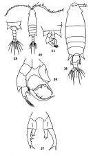 issued from : C.J Shen & S.O.Bai in Acta Zool. Sinica, 1956, 8 (2). [Pl. V, Figs.36-41; as Labidocera bipinnata]. Female: 36, habitus (dorsal view); 37, P5; Male: 38, Th5 and Urosome (dorsal view); 39, P5; 40, habitus (dorsal view); 41, specimen of intersex: Th5 and P5 (ventral view).
|
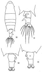 issued from: Q.-c Chen & S.-z. Zhang in Studia Marina Sinica, 1965, 7. [Pl.39, 10-11]. As Labidocera bipinnata. Female (from E China Sea): 10, habitus (dorsal); 11, urosome (ventral); 12, urosome (another specimen), dorsal; 13, urosome (another specimen), ventral.
|
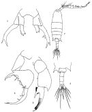 issued from: Q.-c Chen & S.-z. Zhang in Studia Marina Sinica, 1965, 7. [Pl.40, 1-5]. As Labidocera bipinnata. Female: 1, P5 (posterior). Male: 2, habitus (dorsal); 3, postero-lateral angles of the last thoracic segment and urosome (ventral); 4, right P5 (anterior); 5, left P5 (anterior).
|
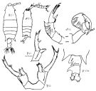 Issued from : K.A. Brodskii in Calanoida of the Far Eastern Seas and Polar Basin of the USSR. Opred. Fauna SSSR, 1950, 35 (Israel Program for Scientific Translations, Jerusalem, 1967) [p.410, Fig.291]. As Labidocera bipinnata. With doubt. Female (from Sea of Japan): habitus (dorsal); AVe, posterior thoracic segment and urosome (ventral); S5, P5; S5 variant, P5 variant. Nota: P5 slightly asymmetrical. Apex of left exopodite with 3 spines, that of right exopodite with 2 spines (3rd spine minute, hardly visible); inner side of exopodite smooth on right leg, with 2 spines on left leg; endopodites 1-segmented, with denticulate apex; spines somewhat variable in number and arrangement. Male: habitus (dorsal); S5, P5.
|
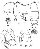 issued from : C.-j. Shen & S.-o. Bai in Acta Zool. sin., 1956, 8 (2). [Pl.V, Figs.36-41]. Female (from Yellow River estuary): 36, habitus (dorsal); 37, P5. Male: 38, last thoracic segment and urosome (dorsal); 39, P5. Intersexued specimen: 40, habitus (dorsal); 41, postero-lateral angles ofcephalothorax and P5. Nota: The postero-lateral angles of the cephalothorx are similar to those of a typical female, but the abdomen is 4-segmented instead of 3, the right A1 is geniculate, P5 is intermediate between the typical female and the typical male, the right leg is generally similar to the left leg of the typical male, the left leg closely resembles the right one of a typical female.
|
 issued from : A. Fleminger, B.H.R. Othman & J.G. Greenwood in J. Plankton Res., 1982, 4 (2). [Fig.4, M-N). Female: M-N, posterior part of prosome and urosome (lateral and dorsal, respectively).
|
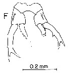 issued from : A. Fleminger, B.H.R. Othman & J.G. Greenwood in J. Plankton Res., 1982, 4 (2). [Fig.5, F). Female: F, P5 (posterior view).
|
 issued from : A. Fleminger, B.H.R. Othman & J.G. Greenwood in J. Plankton Res., 1982, 4 (2). [Fig.6, K-L). Male: K-L, posterior part of prosome and urosomal segments 1-3 (lateral and dorsal, respectively).
|
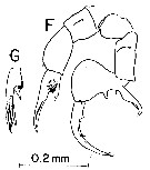 issued from : A. Fleminger, B.H.R. Othman & J.G. Greenwood in J. Plankton Res., 1982, 4 (2). [Fig.7, F-G). Male: F, P5 (posterior view); G, left apical segment (lateral view)..
|
 issued from : A. Fleminger, B.H.R. Othman & J.G. Greenwood in J. Plankton Res., 1982, 4 (2). [Fig.8, F). Male: F, right A1 (dorsal view).
|
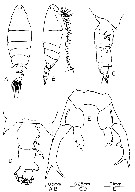 issued from : B.H.R. Othman & T. Toda in Coastal Mar. Sc., 2006, 30 (1). [p.314, Fig.16]. Female (from Sister's Island, Singapore): A-B, habitus (dorsal and lateral, respectively); C-D, posterior part of prosome and urosome (lateral and dorsal, respectively); E, P5. Nota: Prosome to urosome length ratio 3.32 : 1. - Body elogated ending in a single spiniform process, right and left thoracic process symmetrical. - Cephalon and 1st pedigerous somite separated. - Cephalic hooks and small dorsal eye lens present. - A1 symmetrical extends to the genital segment. - Urosome 3-segmented. - Genital segment expanded near midlateral margin shaped of a knob and with 4 spiniform processes along the lateral margin. - Urosomite 2 expanded like a knob at the distal lateral right margin,proximal lateral right margin wuth expanded spiniform process pointing posteriorly. -P5 asymmetrical; right leg slightly longer than left leg.Left exopod similar to pectinata; right exopod with small intercalary denticle between distal spiniform processes, medial margin with 2 denticles
|
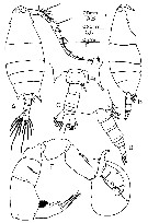 issued from : B.H.R. Othman & T. Toda in Coastal Mar. Sc., 2006, 30 (1). [p.314, Fig.17]. Male (from Sister's Island, Singapore): A-B, habitus (dorsal and lateral, respectively); C-D, posterior part of prosome and urosome (dorsal and lateral, respectively); E, P5. Nota: Prosome to urosome length ratio 3.13 : 1. - Body same as female but cephalic hooks, dorsal eye lens larger than females and contiguous. - Right A1 geniculate villiform teeth on segments 18-21. Last thoracic segment asymmetrical, left corner as in female , right corner similar to that of pectinata except that the denticles between the lateral and medial spiniform processes is more pronounced. - Genital segment with acicular process reaching slightly beyond middle of urosomite 2. - P5 with apical segment of left leg, in general, similar to pectinata, however it differs from it in that here the origin of the spiniform and setiform processes is distal to the tip of spur whereas in pectinata the origin is proximal to the tip; right leg thumb of chelate with pronounced hook at distal end.
|
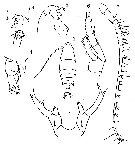 issued from : O. Tanaka in Jap. J. Zool., Tokyo, 1936. [Plate II, figs.1-7]. as Labidocera bipinnata]. Female (from Sagami Bay): 1, habitus (dorsal view); 2, Head (lateral view); 3 last thoracic segment and Urosome (ventral view); 4, the same (dorso-lateral view); 5, right A1; 6, P1; 7, P5. Nota: Head with side hooks. - Rostrum shory and bifid at apex. - Last thoracic segment nearly symmetrical; lateral margins produced into sharp points. - Urosome 3-segmented; the combinede length of the abdomen and caudal rami contained 3.5 times in the total length of cephaloyhorax. - Genital segment asymmetrical, with a prominent process on the right side and 2 setae on the ventral side - The urosomite 2 expands into a wing-shaped projection with a pointed distal end. - Anal segment very short. - Caudal rami asymmetrical, the left one about twice as large as the right and bears a protuberance on the inner margin. - A1 23-segmented, extend about to the end of the genital segment. - A2, mouth appendages and P1 to P4 nearly similar to the other members of this genus. - P5 asymmetrical. The outer margin of exopod with 2 small spines; left exopod furnished with 3 setae on the inner margin, the apex terminates into 3 unequal spines. The apex of the right exopodite ends in 2 spines; the endopodite is differentially denticulated at the apex as in L. pectinata.
|
 issued from : O. Tanaka in Jap. J. Zool., Tokyo, 1936. [Plate II, fig.10]. as Labidocera bipinnata]. Male: 10, habitus (dorsal). Nota: The shape of the head resembles that of the female. - last thoracic segment asymmetrical, the right side is produced into a bifid process, with several small spines on the posterior margin, while the left side is about same as in the female.
|
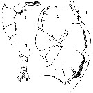 issued from : O. Tanaka in Jap. J. Zool., Tokyo, 1936. [Plate III, figs.1-4]. as Labidocera bipinnata]. Male: 1, Right A1; 2, right P5; 3, left P5; 4, urosome (ventral). Nota: The 18th segment of the clasping A1 has 2 rows of fine teeth on the upper margin, the succeding segment is the longest, with 2 rows of fine teeth. The 22nd segment is produced on the upper margin distally into a long, slightly denticulated spine about as long as the 22nd segment. - The thumb-like process on the proximal end of the outer margin of the 1st segment of the right exopodite is stout and curved; the middle of the outer margin is furnished with a small pointed tooth; the claw-like 2nd segment terminates in 2 spines on the apex. The left exopodite bears 2 spines and 2 setae on the apex, and 1 stout spine on the inner margin, close to the pad of fine hair. Remarks: The male of this species resembles that of L. kröyeri (Brady).
|
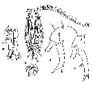 issued from : T. Mori in The Pelagic Copepoda from the neighbouring waters of Japan. S. Shirai ed., Tokyo, 1964 (2nd edit.). [Pl.43, Figs.2, 4, 5, 6]. As Labidocera bipinnata. Female (from 34°18'N, 126°25'E): 2, habitus (dorsal); 4, P5 (left side); 5, P5 (right side); 6, Urosome (ventral). Nota: Head with lateral hooks, but has neither crest nor median hook. - Rostrum symmetrical. - Lateral angles of the last thoracic segment pointed. Urosome asymmetical. - Genital segment produced into process on the right side, and has 2 spine-like projections on the ventral side. - 2nd urosomal somite with a large pointed projection on the right side. Anal segment short. - Caudal rami asymmetrical; the left caudal ramus with a blunt process on the inner margin. - P5 symmetrical; exopodite with 2 outer marginal spines, and terminates into the trifurcate end, often has 2 spines on the middle portion of the inner margin; the endopodite is furnished with many spinules on the terminal and external margins.
|
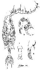 issued from : T. Mori in The Pelagic Copepoda from the neighbouring waters of Japan. S. Shirai ed., Tokyo, 1964 (2nd edit.). [Pl.43, Figs.1, 3, 7, 8]. As Labidocera bipinnata. Male (34°18'N, 126°25'E): 1, habitus (dorsal); 3, P5; 7, urosome (ventral); 8, terminal portion of left P5. NotaThe lateral angles of the last thoracic segment pointed and asymmetrical, the right side bifurcate. - Urosome 5-segmented. - Genital segment with 1 spine on the ventral side. - The succeding segment of the knee-like articulation of the grasping A1 nearly as long as the next segment. - P5 asymmetrical; the thumb of the forceps is curved and pointed. The outer margin of the 1st segment of exopodite of the right leg is nearly straight and has a small blunt tooth.
|
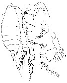 issued from : T. Mori in Zool. Mag. Tokyo, 1929, 41 (486-487). [Pl. X, Figs.1-8]. Male (from Chosen Strait, Korea-Japan): 1, habitus (lateral); 2, posterior angle of left side, last thoracic segment; 3, urosome; 4 (posterior), P4; 5, forehead (ventral); 6, left P5; 7, right P5; 8, left A1. Nota in Mori, 1964, p.95 (from 35°27'30''N, 129°29'E): Head without crest nor median hook and lateral hooks. - The lateral angles of the last thoracic segment are pointed and asymmetrical, the right side is bifurcate, but the left terminates into the simple send. - Genital segment with a dilatation on the right side. - The immediately following segment of the knee-like articulation of the grasping A1 is longer than 2 times as long as the following one. - Right P5 composed of a forceps; the outer margin of the 1st segment of exopodite with a blunt tooth and 1 spine; the terminal claw has 2 spines on the outer margin, and terminates into bifurcate end.
| | | | | Ref. compl.: | | | Madhupratap & Haridas, 1986 (p.105, tab.1); Fleminger, 1986 (p.84, figs. 7, 8, Rem.: geographic vs Wallace's Line); Rezai & al., 2004 (p.489, tab.2, Rem.); Ohtsuka & al., 2008 (p.115, Table 5); Itoh & al., 2011 (p.129, vertical distribution); Beltrao & al., 2011 (p.47, Table 1, density vs time); DiBacco & al., 2012 (p.483, Table S1, ballast water transport); Moon & al., 2012 (p.1, Table 1); Jang M.-C & al., 2012 (p.37, abundance and seasonal distribution); Tachibana & al., 2013 (p.545, Table 1, seasonal change 2006-2008); Suzuki, K.W. & al., 2013 (p.15, Table 2, 3, 4, estuaries, annual occurrence); Seo & al., 2013 (p.448, Table 1, occurrence); Park E.-O. & al., 2013 (p.165, Fig.5, distribution vs stations); Trottet & al., 2017 (p. 7, Table 3: resting stage); Ohtsuka & Nishida, 2017 (p.565, Table 22.1); | | | | NZ: | 5 | | |
|
Carte de distribution de Labidocera rotunda par zones géographiques
|
| | | | | | | | | | Loc: | | | Kenya, E Indian, Straits of Malacca, G. of Thailand, off Bankok, Singapore Harbor, Hong-Kong, China Seas (Bohai Sea, Yellow Sea, East China Sea, South China Sea, Xiamen Harbour, Jiaozhou Bay), Taiwan (SW, E, N), W Korea (Chunsu Bay, Seomjin River estuary, Muan Bay), Korea Strait, Japan Sea, Japan (Ariake Bay, Sagami Bay, Tokyo Bay, Tanabe Bay, Seto Inland Sea, south estuaries : Chikugo, Midori & Kuma Rivers), Amur and Poseta Bays, Okhotsk Sea, E Siberia | | | | N: | 74 ? | | | | Lg.: | | | ? (22) F: 1,16-1,86; M: 1,26-1,79; (91) F: ± 2,25; M: 1,8; ± 2; (120) F: 1,96-1,16; M: 1,62-1,42; (130) F: 1,86-1,16; M: 1,62-1,42; (269) F: 2,26-1,83; M: 2,14-1,58; (290) F: 2,45-2; M: 2-1,9; ? (351) F: 2,01; M: 1,7; (866) M: 1,26-2; (1086) F: 1,97-2,31; M: 1,55-2,02; {F: 1,16-2,45; M: 1,26-2,14} | | | | Rem.: | Labidocera bipinnata Tanaka,1936 (F,M)
Ref.: cf. supra
Ref. compl.: cf. supra
Loc.: G. of Thailand, China Seas, Xiamen Harbour, Taiwan, Yellow Sea, S Korea, Japan, E Siberia, Okhotsk Sea
N: 32
Lg.: (22) M: 1,79-1,26; (91) F: ± 2,25; M: ± 2; (120) F: 1,86-1,16; M: 1,62-1,42; (130) F: 1,86-1,16; M: 1,62-1,42; {F: 1,16-2,25; M: 1,26-2,00}
Rem.: Un doute subsiste sur la synonymie des deux espèces.
Voir aussi les remarques en anglais | | | Dernière mise à jour : 09/12/2020 | |
|
|
 Toute utilisation de ce site pour une publication sera mentionnée avec la référence suivante : Toute utilisation de ce site pour une publication sera mentionnée avec la référence suivante :
Razouls C., Desreumaux N., Kouwenberg J. et de Bovée F., 2005-2026. - Biodiversité des Copépodes planctoniques marins (morphologie, répartition géographique et données biologiques). Sorbonne Université, CNRS. Disponible sur http://copepodes.obs-banyuls.fr [Accédé le 11 février 2026] © copyright 2005-2026 Sorbonne Université, CNRS
|
|
 |
 |


