|
|
 |
Fiche d'espèce de Copépode |
|
|
Calanoida ( Ordre ) |
|
|
|
Epacteriscoidea ( Superfamille ) |
|
|
|
Ridgewayiidae ( Famille ) |
|
|
|
Exumella ( Genre ) |
|
|
| |
Exumella mediterranea Jaume & Boxshall, 1995 (F,M) | |
| | | | | | | Syn.: | Exumella polyarthra : Carola & al., 1995 (p.148, figs.F, st.5) | | | | Ref.: | | | Jaume & Boxshall, 1995 (p.93, figs.F,M); Barthélémy, 1999 a (p.10, fig.); Suarez-Morales & Iliffe, 2005 (p.420-421: Rem.); Vives & Shmeleva, 2007 (p.987, figs.F,M, Rem.); Laakmann & al., 2019 (p.330, fig. 2, 3, phylogenetic relationships) | 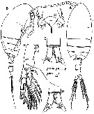 issued from : D. Jaume & G.A. Boxshall in Sarsia, 1995, 80. [p.95, Fig.1]. Female (from Mallorca, Balearic Islands): A-B, habitus (dorsal and lateral, respectively); C, rostrum (fro,tal view); D, anal somite and caudal rami (dorsal); E, idem (ventral); F, P5 (posterior). Nota: Rostral sensory complex composed of 2 raindrop shaped vesicles each opening via a small pore, 2 sensilla, and a pair of minute pores between sensilla. Head and 1st thoracic segment fused (incomplete transverse suture visible dorsally, Th4 and Th5 separated. Th5 segment asymmetrical, showing traces of a dorsal lobe on right side of posterior margin; 2 longitudinal rows of sensilla implanted along both sides of pedigerous somites 2-4. Urosome 3-segmented. Genital double-somite asymmetrical, produced on the right side, with single gonopore opening ventrolaterally on right side, operculum subcircular, 2 sensilla positioned anterodorsally and posteroventrally to operculum. Anal somite bearing striated distal hyaline frill forming 2 big triangular teeth on dorsal margin, and 2 smaller on ventral margin. Caudal rami symmetrical, about 1.7 times as long as wide, armed with 1 short subdistal dorsal seta and 5 distal setae; row of setules along medial margin of each ramus. A1 symmetrical, 27-segmented, not reaching distal end of prosome.
|
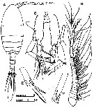
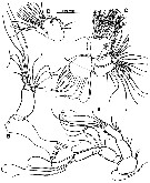 issued from : D. Jaume & G.A. Boxshall in Sarsia, 1995, 80. [p.96, Fig.2]. Female: A, A2; B, Md; C, Mx1; D, Mx1 (detail of basis, exopod and endopod). Nota: A2 with endopod 2-segmented; exopod 7-segmented. Md with coxal gnathobase stout, cutting blade well developed; mandibular palp with endopod reduced, 2-segmented; exopod 4-segmented. Mx1 with well defined praecoxa produced medially into arthrite bearing 10 stout, pectinate spines, submarginal row of 4 setae on ventral side, and isolated dorsal seta; coxa with epipodite armed with 9 setae, and endite with 4; basis fused with exopod and endopod; proximal basal endite discrete, armed with 4 setae; distal incorporated into segment, armed with 5 setae; endopod fully incorporated to basis, formed by 3 indistinctly fused lobes (perhaps remnants of 3 ancestral segments) with setal formula: 2, 4, 5; exopod bearing 10 marginal setae.
|
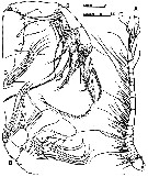 issued from : D. Jaume & G.A. Boxshall in Sarsia, 1995, 80. [p.97, Fig.3]. Female: A, right A1 (ventral); B, Mx2; C, Mx2 (detail of endopod); D, Mxp. Nota: Mx2 comprising completely fused praecoxa and coxa, each armed with 2 endites, allobasis, and 3-segmented endopod; proximal praecoxal endite with 4 long, stout setae, and 1 slender seta; distal with 3 stout setae, 1 longer than others; Both coxal endites armed with 3 setae (2 long and stout, 1 slender and reduced on proximal endite); 3 unequal, with 1 very stout, on distal endite; proximal allobasis endite well developed, swollen, armed with 4 stout unequal setae; distal bearing 3 unequal setae, 1 very reduced in size; free endopod setal formula: 3, 2, 2; 1 of setae on proximal segment very reduced, and another very thick and stout; 1 on distal segment shorter than other. Mxp 6-segmented; syncoxa armed with 7 setae distributed in 4 groups along medial margin; 1, 1, 2, 3; small lobe protruding on distal part of medial margin; row of short setules on distal margin of segment; basis plus fused 1st endopodal segment armed with 3 basal and 2 endopodal setae distally on medial margin; row of short setules along proximal portion of medial margin; free endopod 4-segmented; 1st and nd segments each with 4 setae on distal portion of medial margin; row of short setules along medial margin of 1st segment between 3rd and 4th setae; 4th seta on 2nd segment very long and stout, articulate, bearing specialized ornamentation; endopod segments 3 and 4 reduced; 3rd bearing 4 unequal setae; 4th formed by fusion of ancestral segments 5 and 6 (as indicated by presence of outer margin seta on this segment), with armature of 5 unequal setae
|
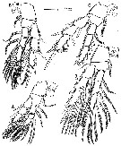 issued from : D. Jaume & G.A. Boxshall in Sarsia, 1995, 80. [p.99, Fig.4]. Female: A-D, P1 to P4 (anterior views).
|
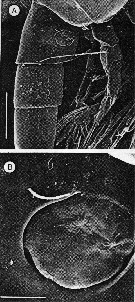 issued from : D. Jaume & G.A. Boxshall in Sarsia, 1995, 80. [p.101, Fig.6 A, B]. Female: A, genital double-somite (right lateral); B, detail of operculum and gonopore. Scale bar: 0.010 mm.
|
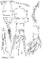
 issued from : D. Jaume & G.A. Boxshall in Sarsia, 1995, 80. [p.100, Fig.5]. Male: A, habitus (dorsal); B, right A1 (ventral); C, P5 (anterior); D, detail of left exopod segment 2 of P5; E, detail of right exopod segment 2 of P5. Nota: Prosome 3.6 times longer than urosome. 5th pedigerous somite symmetrical. Urosome 4-segmented. Genital somite asymmetrical, slightly produced and extended backwards on left side; single gonopore opening ventrolaterally on left side close to posterior margin of segment; operculum not so clearly defined as in female genital double-somite, forming narrow bulge dorsal to gonopore. Anal somite and caudal rami as in female. A1 asymmetrical, left as in female; right geniculate, 24-segmented, with segments II, III and IV, X and XI, and XII, XIII and XIV partially fused. P5 asymmetrical, both biramous; endopod 3-segmented; exopod 2-segmented.
|
 issued from : M. Carola, C. Razouls & J.L. Pretus in Vie Milieu, 1995, 45 (2). As Exumella polyarthra. Female (from Minorca, Balearic Islands): A, habitus (dorsal); B, urosome (ventral); C, urosome and spermatophore (lateral); D, A1; E, P5. Nota : no differences have been observed between the individuals of Minorca and those of Bahamas described by Fosshagen (1970).
|
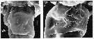 issued from : R.-M. Barthélémy in These Doc. Univ. Provence (Aix-Marseille I), 1999. [Fig.15, E, F]. Female (from La Ciotat, France): E, external ventral view of genital double-somite; F, right lateral position of genital area go = genital operculum; Scale bars: 0,010 mm.
| | | | | Ref. compl.: | | | Giacomo & al., 2005 (p.98); Pansera & al., 2014 (p.221, Table 2, abundance); Belmonte, 2018 (p.273, Table I: Italian zones) | | | | NZ: | 1 | | |
|
Carte de distribution de Exumella mediterranea par zones géographiques
|
| | | | | | 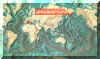 Carte de 1996 Carte de 1996 | |
| | | | Loc: | | | W Medit. (Baleares (Majorca, Minorca), La Ciotat (cave), Sicily (Lake Faro), Messina Strait | | | | N: | 5 | | | | Lg.: | | | (507) F: 1,06; M: 0,94; (511) F: 0,89-0,84; {F: 0,84-1,06; M: 0,94} | | | | Rem.: | La découverte concomitante de cette forme très proche de E. polyarthra , aux Baléares et près de La Ciotat à l'est de Marseille (R. Barthélemy, 1997, comm. pers.) dans des grottes sous-marines, est une illustration de l'isolement d'une population lors de l'ouverture de l'Atlantique. Son maintien pose le problème de l'assèchement plus ou moins important de la Méditerranée au Miocène. Jaume & Boxshall (1995, p.104) ne trouvent aucune differences morphologiques majeures entre les populations de Sardaigne et celles des Baléares. Celles-ci sont isolées par environ 400 km avec des fonds excedant 2000 m. D'autres crustacés dans des excavations sous-marines sont connus en Méditerranée.
Voir aussi les remarques en anglais | | | Dernière mise à jour : 06/02/2020 | |
|
|
 Toute utilisation de ce site pour une publication sera mentionnée avec la référence suivante : Toute utilisation de ce site pour une publication sera mentionnée avec la référence suivante :
Razouls C., Desreumaux N., Kouwenberg J. et de Bovée F., 2005-2025. - Biodiversité des Copépodes planctoniques marins (morphologie, répartition géographique et données biologiques). Sorbonne Université, CNRS. Disponible sur http://copepodes.obs-banyuls.fr [Accédé le 05 juillet 2025] © copyright 2005-2025 Sorbonne Université, CNRS
|
|
|
|


;)
;)
;)
;)
;)
;)
;)

;)
