|
|
 |
Fiche d'espèce de Copépode |
|
|
Calanoida ( Ordre ) |
|
|
|
Epacteriscoidea ( Superfamille ) |
|
|
|
Ridgewayiidae ( Famille ) |
|
|
|
Ridgewayia ( Genre ) |
|
|
| |
Ridgewayia klausruetzleri Ferrari, 1995 (F,M) | |
| | | | | | | Ref.: | | | Ferrari, 1995 (p.182, figs.F,M, juv.); Ferrari & Benforado, 1998 a (p.209, figs. juv., F,M); Ferrari & Dahms, 2007 (p.37, 56, 57, 60, 83, 87, 94, 95, , 96, figs. copepodites I-VI, fig.24, Table VI, XVII, XVIII, XIX, XXII, XXV, fig.30: P1); Figueroa & Hoefel, 2008 (p.145, Table 2, 3, Rem.); | 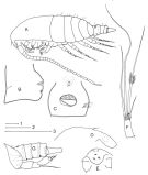 issued from : F.D. Ferrari in Proc. Biol. Soc. Washington, 1995, 108 (2). [p.182, Fig.1]. Female (from NW Atlant.: Belize): A, habitus (lateral left side); B, genital complex (lateral right side); C, idem (ventral); D, forehead and rostrum (lateral); E, forehead and rostrum (ventral); F, caudal ramus (dorsal). Male: G, posterior end of cephalothorax and urosomal somites (lateral left side). Bars: 1 = 0.1 mm (A); 2 = 0.1 mm (G); 3 = 0.1 mm (B-F). Nota: A1 26-segmented. Genital complex slightly asymmetrical (viewed ventrally), several sensilla near left posterolateral corner. Copulatory and oviducal openings separate.
|
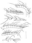 issued from : F.D. Ferrari in Proc. Biol. Soc. Washington, 1995, 108 (2). [p.184, Fig.2]. Female: A, A1 (segments 1-9); B, idem (segments 10-20); C, idem (segments 20-26). Male: D, right A1 (segments 1-10); E, idem (segments 11-18); F, idem (segments 19-21). Bar: 0.1 mm.
|
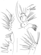 issued from : F.D. Ferrari in Proc. Biol. Soc. Washington, 1995, 108 (2). [p.185, Fig.3]. Female: A, Mx1 (anterior); B, Mx2 (medial lobe 1, posterior); C, Mx2 (posterior); D, Mxp (syncoxa and basis, posterior); E, Mxp (endopod, posterior). Bar: 0.1 mm. Numbers indicate the appearance of endopodal segments during development.
|
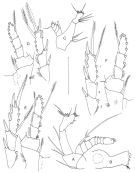 issued from : F.D. Ferrari in Proc. Biol. Soc. Washington, 1995, 108 (2). [p.186, Fig.4]. Female: A, A2; B, Md (mabndibular blade); C, Md (madibular palp); D, P2 (posterior, exopod detached); E, P3 (posterior, exopod detached); F, P4 (posterior, exopod detached). Bar: 0.1 mm. Numbers indicate the appearance of exopodal segments during development.
|
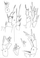 issued from : F.D. Ferrari in Proc. Biol. Soc. Washington, 1995, 108 (2). [p.187, Fig.5]. Female: A, P1 (anterior, exopod detached); B, basis, endopod 2 and 3 of P1 (medial); C, P5 (posterior). Male: D, right P5 and coxa of left P5 (anterior); E, right exopod 1 of P5 (posterior); F, left basis, exopod and endopod of P5 (anterior); G, left exopod 1 and 3 of P5 (anterior). Bar: 0.1 mm.
|
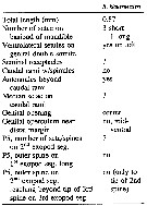 issued from : D.F. Figueroa & K.L. Hoefel in J. Crustacean Biol., 2008, 28 (1). [p.145, Table 2]. Characters female of R. klausrueltzlzri.
|
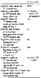 issued from : D.F. Figueroa & K.L. Hoefel in J. Crustacean Biol., 2008, 28 (1). [p.145, Table 3]. Characters male of R. klausrueltzlzri.
|
 Issued from : V.N. Andronov in Russian Acad. Sci. P.P. Shirshov Inst. Oceanol. Atlantic Branch, Kaliningrad, 2014. [p.45, Fig.13, 4]. Ridgewayia klausruetzleri after Ferrari, 1995. Exopod of P1 female.
| | | | | NZ: | 1 | | |
|
Carte de distribution de Ridgewayia klausruetzleri par zones géographiques
|
| | | | Loc: | | | W Atlant. (Belize) | | | | N: | 1 | | | | Lg.: | | | (517) F: 0,9-0,84; M: 0,82-0,77; {F: 0,84-0,90; M: 0,77-0,82} | | | | Rem.: | This species resembles R. marki, but can be separated by different characters: position of the copulatory pore on the genital segment, the shape of the distomedial corner of the 2nd (proximal) endopodal segment of the female P5, and so on (see Ferrari, 1995, p.196). | | | Dernière mise à jour : 20/04/2016 | |
|
|
 Toute utilisation de ce site pour une publication sera mentionnée avec la référence suivante : Toute utilisation de ce site pour une publication sera mentionnée avec la référence suivante :
Razouls C., Desreumaux N., Kouwenberg J. et de Bovée F., 2005-2025. - Biodiversité des Copépodes planctoniques marins (morphologie, répartition géographique et données biologiques). Sorbonne Université, CNRS. Disponible sur http://copepodes.obs-banyuls.fr [Accédé le 30 novembre 2025] © copyright 2005-2025 Sorbonne Université, CNRS
|
|
 |
 |


